Lecture Notes for March 8th: More on Ectoderm and Endoderm

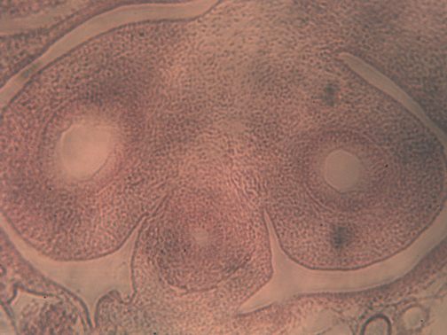
Mouse primordial Germ Cells arriving at the genital ridge.
This is an original photograph of Primordial Germ Cells that was taken by a student in this course many years ago who collaborated on research with Dr. Mitch Eddy at the NIEHS in the Research Triangle Park.
We were looking for evidence of chemotaxis, but couldn't prove whether the PGCs were being guided by chemotaxis or haptotaxis.


Neural Crest Ectoderm:
Sensory nerve, dorsal root ganglia (one per somite).
(neural segmentation Induced (is that the right word?) by earlier segmentation of somites)
(This is an interesting version of geometric arrangements of nerve cells & axons
(being somehow controlled by locations where somites previously had existed)
(Is this caused by chemical signals, or by physical forces?)
Postganglionic autonomic nerves (Preganglionic autonomic nerves are from neural tube)
Melanocytes, and other mesenchymal pigment cells.
Schwann Cells (but not oligodendrocytes, which are derived from neural tube) [Multiple sclerosis is an immune attack on oligodendrocytes but not Schwann cells?!]
Facial Skeleton. (Cell types that would be mesodermal in any other part of the body!) [The explanation of this is unknown - and debated]
Stomodeum (is Ectoderm, not endoderm)
Ameloblasts: produce outer enamel layer of teeth. (These begin as epithelial invaginations into what will become the gums.)
Rathke's Pocket: an upward epithelial invagination from the roof of the mouth. Develops into the Anterior Pituitary Gland (Fuses with a downward epithelial invagination from the floor of the mid-brain, which differentiates into the Posterior Pituitary Gland.)
In mammals, the roof of the mouth becomes closed off by two palatal shelves, which swing toward each other like two doors, and fuse to form the palate.
Note that birds and most reptiles do not have such a structure. (Alligators are a special exception)
An all-too-common human birth defect is "cleft palate", which results if the palatal shelves do not fuse with each other.
There is no obvious boundary between the oral ectoderm versus the endoderm (the oral ectoderm is the Stomodeum vs. the Archenteron forms the Endoderm). But a rare birth defect consists of a failure of these epithelia to fuse. The result is continued existence of an Oropharyngeal Membrane. (Which blocks the throat, and has to be surgically removed at birth)
[I would have expected some discontinuity, such as a change in the epithelial structure, would continue to exist during life. But nothing is visible.]
Endoderm (A series of organs form as epithelial invaginations of endoderm)
4 pharyngeal pouches (and 4 actual gill slits, which then close). These begin as epithelial invaginations in the sides of the endodermal tube.
"Cervical Fistulas" are birth defects in which open channels continue to exist, and don't close. These can be surgically repaired.
[How a surgeon removes a hole is metaphysically worth wondering about!]
First Pharyngeal Pouch --> Eustachian Tube and external ear opening.
Second Pharyngeal Pouch --> Tonsils
Third Pharyngeal Pouch --> Parathyroid Gland and Thymus
Fourth Pharyngeal Pouch --> Parathyroid Gland and Thymus
Certain birth defects in humans consist of under-development of 3rd & 4th "gill slits". The symptoms were very puzzling: reduced immunity and lack of control of calcium and phosphate. Eventually, an MD remembered what develops from the 3rd and 4th pharyngeal pouches. Until then, scientists interpreted this birth defect as evidence that immunity might be controlled by calcium and/or phosphate concentrations, or the reverse.
Really, the connection is that both are controlled by organs that happen to develop from the same early embryonic structures. More discoveries like this are waiting for you be the person who figures out the explanation.
Thyroid gland: Develops as epithelial invaginations in the floor of the anterior endoderm.
Separates off from the esophagus, and becomes a bunch of hollow sacs
Thyroxine is formed by reactions between tyrosine and iodine
in extracellular gels inside these epithelial sacs (instead of inside the cells)
This evolved from a groove-like down folding in the throats of sea squirts, called the endostyle, which secretes a gel used in filter-feeding.
Salivary Glands develop as epithelial invaginations in the wall of the endodermal tube. (4 out of the 6 salivary glands do, anyway. One pair invaginate from the stomodeum wall.
Lungs develop as epithelial invaginations in the wall of the endodermal tube.

Swim bladder develops as an epithelial invagination in the wall of the endodermal tube.
Pancreas gland develops as an epithelial invagination in the wall of the endodermal tube.
Liver develops as an epithelial invagination in the wall of the endodermal tube.
The cloaca develops as an epithelial out folding in the posterior the endodermal tube.
In Mammals (but not. birds, reptiles, amphibians of fish) the cloaca becomes split into the bladder and the rectum.
We have cloacas when we are embryos, but these become physically separated into the anterior part (which becomes the bladder) and the posterior part which becomes the rectum.
****************************************************************************
QUESTIONS TO THINK ABOUT AND DISCUSS DURING SPRING BREAK:
a) How many birth defects result from failure of embryonic structures to fuse with each other?
b) What heart defects could you include in this category?
d) Please try to invent as many possible birth defects as you can imagine, that would be caused by failure of fusion between sheets of embryonic cells whose fusion is part of normal development.
(Please note that many of your imaginary birth defects actually occur, but I just didn't include them.)
d) How many examples can you remember in which important organs develop as epithelial invaginations? Can you think of ten examples? Twenty?
e) How many examples can you think of in which epithelial folds become the boundaries between cells that differentiate to become very different cell types. (Dozens of examples occur).
****************************************************************************
REVIEW: I) Hairs, feathers, finger-nails and (reptile) scales begin as epithelial invaginations in the somatic ectoderm.
II) Teeth (enamel) and anterior pituitary develop from epithelial invaginations in the sides and roof of the stomodeal ectoderm.
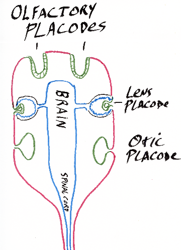
III) The nostrils and the sensory part of the nose, the lens of eye, inner ear and the lateral line organ develop as thickenings (placodes) and large invaginations of head and neck somatic ectoderm.
IV) Liver, pancreas, lungs, swim bladder, salivary glands, endostyle, and thyroid gland develop as epithelial invaginations in the wall or floor of the epithelial tube that began as the archenteron.
****************************************************************************
HAVE A HAPPY SPRING VACATION.