Biology 441, Spring 2013: Ectoderm, placodes
Lecture notes for Wednesday, March 6
Ectodermal cells are sub-divided into three categories

(#1) Somatic ectoderm
-
Epidermis layer of skin,
and fingernails, hair, feathers, scales (of reptiles & birds)
(teleost scales are mesodermal, however)
Lens of eye,
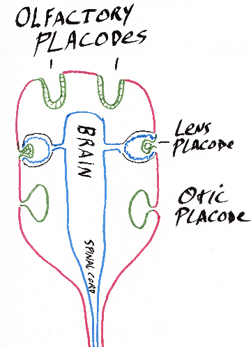
Inner ear [cochlea etc.] (formed from Otic placode)
Olfactory placode -> Sensory nerves of nose
Other Placodes which form nerves in head area.
Other Placodes that form the Lateral Line of fish & amphibia
___________________________________________________________________
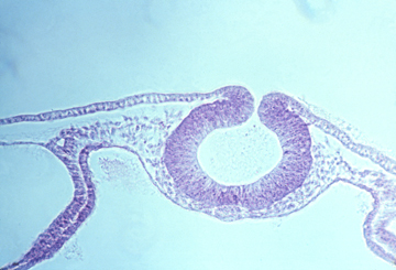
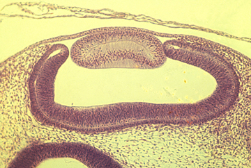
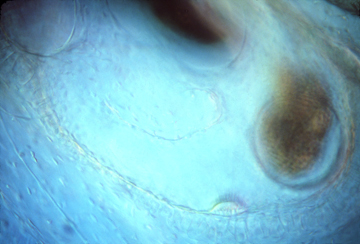
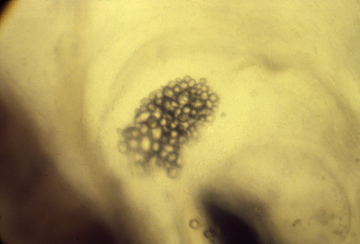
Also formed by out-foldings from the sides of the brain are the optic cups, the outermost edges of which bend back inward to form the neural retina. The part of the optic cup just inward from the neural retina differentiates into a thin epithelium full of melanin pigment granules, called the pigmented retina.
Small areas of somatic ectoderm are induced by the optic cups to fold inward and differentiate into the lens of the eye. Lens cells elongate very much.
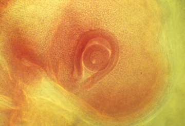
Eye cup with lens
___________________________________________________________________
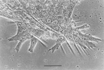
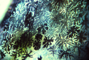
The Lateral Line System is a set of grooves and tunnels just under the skin in the head and down the sides of fish, that are sensory organs for water flow, and use the same kind of Neuromast Cells ('hair' cells) used in semi-circular canals. Frogs and salamanders also have lateral line systems. But reptiles, birds and mammals never develop lateral line systems.
Neuromast cells have special filopodia that detect even very small bending forces, which alter the frequency of nerve impulses sent to the brain. These filopodia are supported by fibers of the muscle protein actin (and are much smaller than hairs on your head, to which they are only similar in shape.
&&&&&&&&&&&&&&&&&&&&&&&&&&&&&&&&&&&&&&&&
While we are on the subject of neuromast cells and the inner ear:
Inertia is detected by water flow in the 3 semi-circular canals bending filopodia of sets of neuromast cells located in parts of these canals.
Gravity is detected by the weight of two (mutually perpendicular) sets off otoliths
which are crystals of calcium carbonate attached to the filopodia of other hair cells.
Sound is detected by long rows of neuromast cells whose filopodia get vibrated against
by special flaps of tissue, arranged so that different frequencies of sound will mostly
stimulate different neuromast cells (because of combinations of different resonances.
In mammals, the sound-detecting part of the inner ear is lengthened and coiled to form a structure named the cochlea. Only mammals have a cochlea, but all vertebrates have part of their inner ears specialized to detect sound vibrations. Frog inner ears tend to be tuned to resonate only to certain frequencies, I have read.
__________________________________________________________________
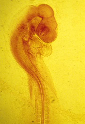
In the mechanical sense, expansion of vertebrate brains is equivalent to inflation of several closely spaces balloons. Cells pump water into the neurocoel, creating enough water pressure to expand the anterior part of the neural tube. The fore-brain, mid-brain andhind-brain are areas which stretch more than the part of the brain between and behind them. Eventually, there is much more cell growth in these areas; But at first, they are thin-walled, water-inflated & balloon-like
Experiments have shown that the brain won't expand if the fluid pressure is released. The eye-ball also depends on hydraulic expansion for its enlargement. (and won't enlarge if punctured)
_________________________________________________________________
(#3) Neural crest ectoderm (sometimes called "ectomesenchyme")
-
segmental sensory nerves (connected to spinal cord) (NOT optic nerve, etc.)
postganglionic autonomic nerves (including the medulla part of the adrenal gland)
Pigment cells of skin ("melanocytes" in Mammals) (plus amphibia, fish, birds & reptiles also have other pigments than melanin and the cells that made these other pigments also develop from neural crest)
-
Schwann cells (that wrap their plasma membranes around nerve axons
(in the peripheral nervous system, to form the myelin sheath.)
-
Facial skeleton (in the anterior part of the head, the cartilages and [probably?] other tissues that are formed by mesoderm in other parts of the body)
It is a big puzzle why neural crest should form skeletal tissues.
Odontoblasts which are special mesenchymal cells that secrete the inner, dentin layer of the teeth.
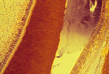
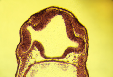
Thin section through tooth development on a mouse embryo.
Enamel is stained purple; dentin is stained dark red
-
#3½ ?) Stomodeum (oral cavity cells, also considered to be ectodermal,
rather than endodermal! Although this may have been more an arbitrary decision than a fact based on any particular evidence, I suspect.)
Lining of mouth (and also nasal cavity) (posterior to oro-pharyngeal membrane)
The palate is the roof of your mouth (but not in birds, reptiles, amphibia) The palate is formed by fusion of right and left palatal shelves.
Enamel (outside layer of teeth: secreted by ameloblasts)
Rathke's pocket -> Anterior Pituitary Gland
(That secretes FSH, LH, etc.)
At least one of the three pairs of salivary glands