Lecture notes for Friday, February 23, part two
Stomodeum, Endoderm, and Digestive Tract
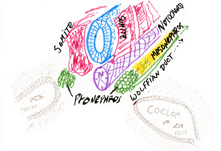
The most anterior end of the digestive tract is formed by an infolding of the ectoderm called the stomodeum.
The mouth:
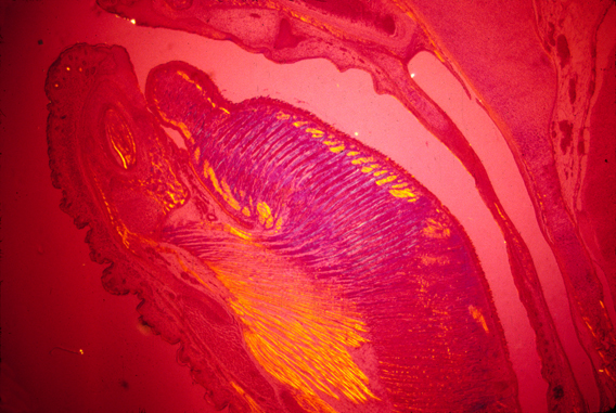
mouse tongue seen with polarized light
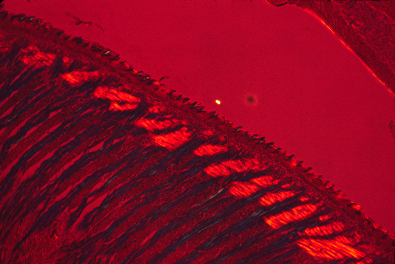
higher magnification showing orientation of muscles in perpendicular directions
The outer layer of teeth (Ameloblasts --> Enamel)

tooth rudiments
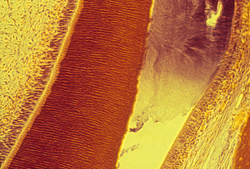
dentin and enamel
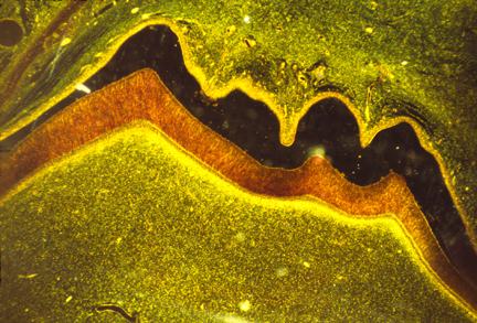
section of a molar
The dark area is empty space, where the enamel had been but broke off
during preparation of the slide. The orange area is the dentin.
pharyngeal pouches:
-
First Pharyngeal Pouch --> Eustachian Tubes
Second Pharyngeal Pouch --> Tonsils
Third Pharyngeal Pouch --> Thymus and Parathyroid Gland
Fourth Pharyngeal Pouch --> Thymus and Parathyroid Gland
Palatal shelves (which are only in mammals, not birds, not reptiles,
not amphibia or fish) separate the oral cavity from the nasal cavity, very much
like a pair of doors closing.
(When the palatal shelves don't fuse, the result is a birth defect called "Cleft Palate")
One of the 3 pairs of salivary glands and the anterior pituitary gland are stomodeal ectoderm.
The rest of the digestive tract develops from endoderm.
The boundary between stomodeum and endoderm is in the back of the throat.
Somewhat surprisingly, there is no detectable change in the tissue at this boundary.
(Not even a historical marker, like the one at Promontory Point, Utah, where transcontinental railways connected)
But a rare birth defect occurs in which the throat is blocked by the "Oro-pharyngeal Membrane" (which results from the endoderm and stomodeum not breaking through, and not creating an opening).
What organs develop from the endoderm? (cells that line the archenteron)
Two of the three pairs of Salivary Glands,
the Thyroid Gland,
the Lungs,
the Pancreas,
the Liver,
the Bladder and Rectum form as outfoldings of the endodermal tube
but only mammals separate the rectum from the bladder:
Birds, reptiles, amphibians, and all kinds of fish have a cloaca
(Latin word for "sewer") into which both the intestine and the Wolffian Duct connect)
(Only) in mammals (but not in birds, reptiles, amphibians and fish), the cloaca becomes separated into
the dorsal rectum
the ventral bladder
But mammal embryos have a cloaca, before this separation occurs.
(Notice the coincidental analogy of the separation of the oral and nasal cavities by the palate.)
(Also notice that these separations occur in mammals, but no other orders of vertebrates.)
One concluding fact:
Amphibians develop the anus from the blastopore (or at least a re-opened hole near where the blastopore was).
Reptiles, birds and mammal embryos don't have blastopores, and the rear end of our digestive tract develops from an infolding called the proctodeum (which is analogous to the stomodeum.)
How fish create this opening, I don't know and would be grateful if someone finds out and tells me.
And a few more facts:
Many organs develop by active folding of epithelial cell sheets:
Examples:
Teeth
Salivary glands
Thyroid glands
Lungs
Pancreas
Liver
(Sample question for the next exam: list 5 organs with gland-like morphology that develop as out-pocketings of the walls of the endoderm)
Differentiation of each of these organs is stimulated by adjacent mesodermal mesenchyme.
-
* They won't develop if their mesenchyme is removed early in development.
* But they can develop if a millipore filter is inserted between the epithelium
and the mesenchyme.
* Sometimes lung epithelium can be induced to differentiate into pancreas, etc. or the reverse, by grafting mesenchyme from one part of the endoderm to another.
* If you see a book, article, or meeting about "Epithelio-Mesenchymal Interactions", it's these sorts of phenomena they will mostly be concerned with.
For example: Teeth
other examples: Scales, Feathers, Hair
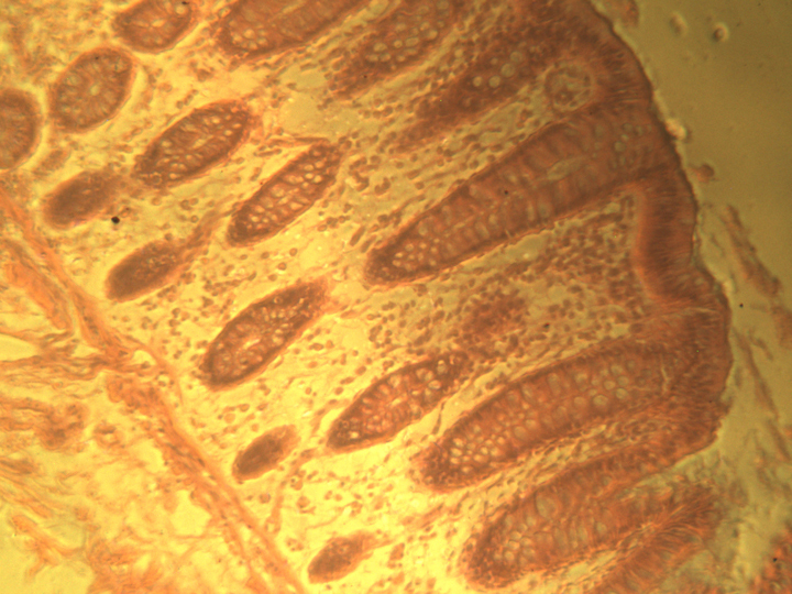
section of large intestine, showing infoldings with mitotic cells