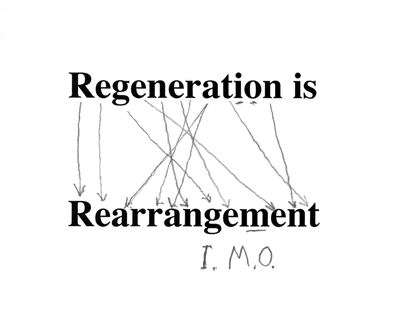
Lecture notes for March 6, 2019
What can we learn about normal limb development from the experimental results (by which I mean Triple Branching) of grafting tip ends of limb buds that have reversed asymmetry?
Learn that regeneration can be different from filling in missing parts?
When tissues are grafted next to something different from their normal neighbors they sometimes respond by filling in whatever anatomy should normally be located between the grafted structure and whatever you grafted next to it.
Imagine Hogwarts students who like to sit together in this order:
Alice_Bob_Clara_Don_Emily_Fred_Georgia_Henry
If Clara, Don, Emily were missing at the breakfast table,
Alice_Bob_-----------------------_Fred_Georgia_Henry?
then Bob and Fred magically create replacements:
Alice_Bob_Clara_Don_Emily_Fred_Georgia_Henry
Suppose they do this by splitting off tissues from Bob and Fred, and converting these tissues into replicas of Clara, Don and Emily.
Next, imagine the result of seating three in reversed order:
Alice_Bob_ Emily_Don_Clara_Fred_Georgia_Henry
_____________________________________________________?
Maybe Bob and Emily don't want to sit next to each other, and neither do Clara and Fred.
How can they use their magic powers to restore normal neighbors?
Bob creates a new Clara! Fred creates a new Emily! What else?
Clara creates a new Don between herself and Emily...
which results in having two Dons! And what else?
ABCDEFG ABEDCFG?
ABcdEDCdeFG?
Everybody is next to their normal neighbors, sometimes by creating extra copies of those neighbors.
Only crazy people could imagine such events!
Except it's what "reverse-grafted" limb buds really do.
______________________________________________?
Surely there are important lessons to be learned about normal interactions between developing tissues.
What are those lessons?
Tissues don't know where they are located, but tissues do know what they are supposed to be next to.
If tissues are put next to "wrong" neighbors, sometimes they react by creating new copies of their correct (missing) neighbors.
Even when this results in producing two, three or even four duplicate copies of their legs or wings.
(However crazy this may seem)
___________________________________________________
Surely Hox genes are involved?
Have you ever heard of the phenomenon of co-linearity of transcription of Hox genes?
The locations in the body at which different hox genes are transcribed have the same relative geometric locations as the positions on the chromosomes where the DNA coding for each different hox gene is located.
Hox#1, Hox#2, Hox#3, Hox#4, Hox#5, Hox#6,
Head, neck, chest, belly, hips, tail
But I over-simplify.
Mammals have four clusters of hox genes, (called A, B, C, and D) with each cluster located on a different chromosome, with up to thirteen variants in each of these four families ….D9, D10, D11, D12, D13 and it's the anterior borders of where each variant hox gene is transcribed that correlates with genetic map locations on the chromosomes.
Nevertheless, (almost) all multicellular animals have this same "co-linearity" of map locations of hox genes on chromosomes correlated with the anterior borders of locations in the body where each hox gene is transcribed, and also correlated with the times when those particular genes are transcribed.
This spatial correlation between map location and anatomical location was first discovered in Drosophila homeotic genes. Even though vertebrates don't have homeotic mutations (such as could cause an extra pair of legs to develop where the arms should be) they do have lots of proteins with nearly the same 61 amino acid sequences by which homeotic genes bind to DNA.
Why isn't more research being focused on this?
How many students in this course have already been taught about hox gene co-linearity?
____________________________________________________
Int J Dev Biol. 2015;59(4-6):159-70. doi: 10.1387/ijdb.150223sg. The significance of Hox gene collinearity. Gaunt SJ1.
Published in 2015 - Cited by 17 QUOTE: … Spatial collinearity (correspondence between ordering of Hox genes along the chromosome and their expression patterns along the head-tail axis) has been conserved in many animal groups …. It is not known why the Hox cluster evolved with spatial collinearity. Four models are discussed. These vary in the significance they place upon Hox chromatin structure, and also on whether they propose that collinearity is primarily concerned with establishment or maintenance of Hox expression. Published proposals to explain spatial collinearity, which invoke enhancer sharing, chromatin closing or chromatin opening, are either problematic or can offer only partial explanations. In an alternative proposal it is suggested here that spatial collinearity evolved principally to maximise physical segregation, and thereby minimize incidence of boundaries, between active and inactive genes within the Hox cluster. This is to minimise erroneous transfer of transcriptional activity, or inactivity, between adjacent Hox genes.
___________________________________________________________?
A hypothetical explanation for co-linearity
First imagine that each cell can detect which Hox gene proteins are being made in themselves, and also that they can detect which Hox gene proteins are being made in neighboring cells.
Second, suppose that cells react strongly to differences between their own Hox proteins and those made by adjacent cells.
Suppose they react in ways analogous to our Hogwarts students Alice, Bob, Clara, Don etc. Specifically, suppose that a cell makes Hox A5, but next-door cells make Hox A7; that is a difference of two! This annoys the cells; being next to cells making Hox A6 would have been tolerable, because that is just a difference of one; not so bad.
But differences of two or more is too different! Something must be done to fix such an outrageous difference, of two or more units. Fixing the situation is easily done: Some cells along the border between A5 and A7 need to change which combination of Hox genes they transcribe, so as to become HoxA6 cells.
An extreme case would be to start with a sheet of many HoxA1 cells with some HoxA13 cells along one side. This discontinuity would stimulate a succession of conversions, the net result of which would be a smooth gradation from HoxA1 cells to HoxA2 cells to HoxA3 cells, and so on.
"The Parable of the Near-Sighted Conformist"!
Scientists would confidently conclude that the spatial gradation of HoxA1, 2, 3, 4, 5, etc. must be a response to a diffusion gradient of some one chemical that can diffuse from cell to cell, and which stimulated transcription of different HoxA genes depending on the local concentration of the diffusing substance. ?
In conclusion, lets try to invent some kind of experiment capable of testing which kind of mechanism causes the co-linearity phenomenon. What if we cut blocks of tissue out of an embryo, rotated their Anterior-Posterior axis by 180 degrees and grafted them back into the embryo with axes reversed. Will the boundaries develop mirror-image reversed self duplications, like those that result when distal ends of limb buds are grafted with reversed polarity? ?
Continuing with limb regeneration in salamanders
I) Newts and many other kinds of salamanders can regenerate their legs.No other vertebrates can do this, except partial regeneration of limbs in frog tadpoles before metamorphosis.
Humans can sometimes regenerate the last digit of fingers and toes, IF the wound is NOT stitched up (which is standard practice).
Regenerating newt legs "re-grow" all the bones (i.e. cartilages), muscles, tendons, blood vessels, connective tissue and skin.
In the stump of the cut limb, cartilage, muscle and other cell type become indistinguishable, "dedifferentiating" back to what looks like their early embryonic state, forming a mass of cells called a blastema, in which rapid cell growth and mitosis replaces the lost cells.
Skeletal muscle cells, which are syncytial, subdivide back to individual cells before resuming mitosis.
It would be interesting to know whether this subdivision of multi-nucleate muscle cells
has the same contractile-ring mechanism as the ordinary cytokinesis that follow mitosis.
The myoblasts then undergo many cycles of mitotic cell divisions, and fuse back together.
II) Grafting of cartilage and muscle labeled with radioactive labeled DNA, and also the use of genetic markers like nucleolus number, consistently show that each cell remains "loyal" to their original differentiated cell type.
In other words the dividing cells in the blastema that had been cartilage cells
re-differentiate only into cartilage,
& the descendants of muscle cells only become muscle.
Most experimenters had not expected this, which make the results even more persuasive.
It also contradicts expectations that regeneration should have the same basic mechanism as the original formation of limbs, except if the original development was by sorting out of cells whose differentiation had already been decided, which nobody considers possible.
Limb regeneration is not thought of as a special case of sorting out by differentiated cells, I guess because there is no deliberate dissociation into single or randomly-mixed cells.
Nevertheless, consider that cartilage cells from which the humerus had previously been constructed separate into what seem to be undifferentiated cells, and then re-differentiate as the radius, ulna digits, wrist bones, as well as replacements for the lost parts of the humerus.
Furthermore, consider that the many muscles of the regenerated leg are made of cells that had been parts of different muscles of the upper limb.
Likewise: skin cells, blood vessels, connective tissue & nerves.
All are replaced by descendants of the same kinds of the cells in the stump of the cut limb.

III) Salamanders are the only vertebrates whose limb buds do not form an apical ectodermal ridge.
However, during regeneration of salamander, the stumps develop an epithelial thickening which is well-formed, but not elongated in the A-P axis (unlike the AER)
I can't figure out how to interpret this combinations of odd facts, but it must mean something.
IV) If you cut off just the "hand" part of a newt leg, then just that part will regenerate.
If you cut the leg off at the elbow or knee, then just the missing part will regenerate.
If you cut off the whole leg, than all the parts of will regenerate.
BUT: If you cut off just the "hand" part, and then paint the cut surface with retinoic acid, then a whole new leg will regenerate, starting over at the shoulder, and forming an extra elbow, etc.
AND: Newts can regenerate their tails.
BUT if you cut off the tail, and paint the cut surface with retinoic acid, guess what regenerates instead!
The more of a newt leg you cut off, the faster tissues regenerate; So that the total time to completion of regeneration is about the same when you cut off the whole leg as when you cut off just the tip.
The Nardi-Stocum Phenomenon of limb regeneration.
Displacement of grafted limb tissue along its proximo-distal axisNardi and Stocum discovered the amazing fact that if you cut off one salamander leg and graft it onto the side of another salamander leg, the graft will move along the other leg until wrist is juxtaposed to wrist, or elbow moves next to elbow, or shoulder next to shoulder. Grafts move until like is next to like.
When elbow cells get grafted to the hand (or to the shoulder) the grafted cells somehow move back to the elbow position.
Also: Hand cells get engulfed by elbow cells,
elbow cells get engulfed by shoulder cells. etc.
Similar to the engulfment of mesoderm cells by ectoderm, engulfment of heart by liver.
From which N&S concluded the cause is what Malcolm Steinberg had hypothesized,
which is thermodynamic (surface tension-like) maximization of cell-cell adhesions. [This will be discussed in more detail in a later lecture.]
Retinoic acid causes big changes in behaviors of embryonic tissues:
"Retinoic acid coordinately proximalizes regenerate patterns and blastema
differential affinity in axolotl limbs":
K Crawford & DL Stocum, Development 1988 cited by 93
"Use of retinoids to analyze the cellular basis of positionl memory in regenerating amphibian limbs"
DL Stocum and K Crawford, Biochemistry and Cell Biology 1987 cited by 51
What is wrong with the following argument?
Tissue engulfment looks like liquid surface tension: Surface tension is caused
by attractive forces pulling molecules toward each other; therefore tissue engulfment
must be driven by maximization of cell-cell adhesion:
& cell adhesion should be measurable by resistance of cell aggregates to flattening.
When Nardi writes "differential affinity", he means cell rearrangement behavior, partly because he regards Steinberg's thermodynamic theory as being proven fact, but also to avoid alternatives like: "That business with the bunches of cells engulfing each other."
The less people understand about thermodynamics, the less they doubt such theories.
(A reason for biologists to take Physical Chemistry is future protection from bluffing.)
Imagine if someone claims "Quantum theory proves Warburg's theory about the cause of cancer"
How can you argue with them? If you ask "How can you prove that?", what will they say?
My point is that even the best scientists can have trouble knowing what evidence proves.
All interpretations of data are attempts to fit observations to expectations, theories & guesses.
______________________________________________________________
All cases where cells gravitate back toward their original spatial arrangement, especially when the same end results can be reached by 2 or 3 or more pathways, get interpreted by many as thermodynamic minimization of free energy.
An alternative explanation is homeostasis by negative feedback cycles.
Imagine if some persuasive person had argued that constant body temperature is caused by
minimization of thermodynamic free energy.
Is this analogous to cell sorting? Nardi and Stocum believe that it is. They believe legs have gradients of cell-cell adhesiveness, with maximum adhesiveness at the hand or foot. Can you figure out what evidence could detect differences of adhesiveness? Could gradients of contractile tension produce the same movement of grafts? Would such knowledge help cause regeneration of legs.
In what sense do grafted wrists sort out? They tried to explain this phenomenon in terms of Steinberg's Differential Adhesion Hypothesis, which we will cover in a later lecture.