-
Notochord
Somites (each of which itself subdivides into 4 parts)
Intermediate Mesoderm (kidneys & male sex ducts)
Lateral Plate mesoderm (coelom, gonads, heart, female sex ducts)

Spatially-periodic structures whose locations are caused by where somites form:
Segmental structures that are ectodermal:
-
Dorsal root ganglia (aggregations of neural crest cells that differentiate into sensory nerves)
Motor nerve bundles
"Dermatomes" in the medical sense. Areas of skin that are innervated by each different segmental sensory nerve.
(I have gotten the impression that motor nerve innervation patterns also are restricted
to analogous bands, like dermatomes)
-
Arteries that branch from the aorta

Tail of a living tadpole. The broad line just below the center
is the notochord, and you can see that it is filled with highly vacuolated
cells. The neural tube is above it. You can also see pigment cells and
mesenchymal cells scattered around.
Somite Formation
At first, there are continuous columns of "paraxial" mesoderm.

The drawing above shows the geometrical rearrangements that some scientists have reported to cause somite segmentation in different classes of chordates.
Top: Amphioxus, Middle: bird and mammal, Bottom: frogs. Anterior is to right sid
Then these columns spontaneously split apart, one pair at a time.
As if a quonset hut were to split into a row of igloos.
Or as if a long piece of french bread were to slice itself.

Longitudinal section of notochord and somites in a developing frog embryo

Somites forming in a fixed whole mount chicken embryo. Anterior to left.

Somites forming in a living chicken embryo. Anterior to left.
The physical mechanism of splitting apart has now been discovered to be stimulation of decreased cell-cell adhesions by specific binding of ephrin proteins.
Ephrins stimulate cells to break their adhesions to each other, and guide optic nerve fibers to their map-like patterns of connections in the roof of the mid-brain.
Some textbooks say that ephrins cause cell to repel each other, but really they induce detachment of adhesions: Cell traction pulls each side in a tug of war, therefore weakening of cell adhesions causes them to crawl directionally away from the weakened adhesions.
In scanning EM photos, "somitomeres" can be seen, where a somite is going to form, apparently as the first stages of somite formation.
In most species, somites start as hollow epithelial balls, and conversion of cells from being mesenchymal to epithelial therefore seems to be part or the process of separation. (These epithelial cells seem to have basal surfaces outward.)
For most species, the same number of somites forms in each embryo.
(the number is 32-34 in humans, 50 in chicks, 65 in mice (many in tail),
more than 500 in some snakes, and varies with temperature in fish!
You form as many vertebrae as your embryo had formed pairs of somites.
Each somite subdivides into four parts:
The dermatome --> cells form the inner layer of skin (dermis)
The myotome ---> all the skeletal muscle cells of the body
The anterior sclerotome --> the posterior half of a vertebra!
The posterior sclerotome --> the anterior half of a vertebra!
Not a typographical error! Each somite gives cells to 2 vertebrae
And each vertebra develops from cells of two adjacent somites.
That way muscles run from one vertebra to the next,
and motor and sensory nerves come out between vertebrae.

Somites only temporarily exist in the embryo:
but their spacing controls future segmentation of many tissues;
vertebrae, ribs, arterial branches
and also sensory ganglia (that develop from neural crest!!)
and also motor nerve bundles from the spinal cord.
Incidentally, to Neurologists a "dermatome" means the stripe-shaped areas of skin and muscles that are innervated by sensory and motor nerves from each segmental spinal nerve. In the disease "Shingles", stripe-shaped zones of blisters form on the skin (looking like shingles on a roof) because rows of blisters form on the skin of different dermatomes.
Neurologists have a method for mapping paralysis caused by slipped disks, by inserting electrodes under the skin and firing one set of nerves at a time. This method hurts quite a bit.

Three somites:
Myotomes in a salamander
Polarization microscopy shows that myotomes develop muscle fibers very early.
Alternating anterior and posterior sclerotomes
Nerve ganglia form only in the anterior sclerotomes. Also notice that small arteries run just below (behind?) each of these ganglia.
Although somites somehow cause the locations of these nerve ganglia and the arteries branching from the aorta,
neither the nerves or arteries develop from somite cells.
Surgical transplantations of salamander somites (5 from the tail replacing 3 from the trunk) prove
that anatomical segmentation is caused by somites themselves,
NOT as additional effects of what-ever signals cause somites to form.
Longitudinal section of 3 somites, that goes right through the myotome
Notice the bundles of nerve fibers located just to the right (toward the midline of the embryo) of each of these three myotomes.
*********************************************************************
Intermediate mesoderm differentiates to form
kidneys, kidney ducts, and male sex ducts.
The most anterior intermediate mesoderm forms
the pronephros ("head kidney") (one on each side).
Pronephros
From each pronephros extends a pronephric duct.
Intermediate mesoderm tubules
In amphibia all the rest of the intermediate mesoderm differentiates into a pair (right & left) of adult kidneys, which use the pronephric duct to carry urine, and to also carry sperm in males.
But in mammals, birds and reptiles, the adult kidney develops from the most posterior parts of the intermediate mesoderm.
These adult kidneys are called the metanephros,
and each one has a special duct, called a ureter.
While we are embryos, we use a separate kidney, that differentiates from the middle parts of the intermediate mesoderm. This is called the mesonephros.
In contrast, you form one single heart (normally),
Imagine re-building a lawn-mower motor into a 4-cycle car engine,
********************************************************************************
Lateral plate mesoderm splits into two layers:
The female sex ducts (Oviducts; Fallopian tubules, Uterus)
differentiate from outfoldings in the coelomic wall.
In Reptiles, Birds and Mammals,
*************************************************
The heart also develops from the extreme right and left sides of the lateral plate mesoderm;
In reptiles and birds, the lateral plate spreads out across the yolk, & completely surrounding the yolk, so as to form the yolk sac.
Embryos of reptiles and birds form their hearts where the right and left lateral plate mesoderm sheets come together, slightly in front of (!) where the head is forming.
Later in development, digestion of yolk causes the yolk sac to get progressively smaller, this shifts the location of the heart in a posterior direction, bringing it into the chest of the embryo.
If pieces of plastic or other physical barriers were inserted where the heart normally forms, so as to block the right and left sheets of lateral plate mesoderm from coming together !!????
What would you expect would happen?
Answer:
#5) Two complete, normal hearts develop
{But they are usually mirror images of each other!}
This is one of several examples in which parts of embryos can be kept apart, or mechanically split.
Another example is that if you cut a limb bud in two;
Or (also like early embryos) you can graft two early limb buds together, and often it will develop into a single large leg, or wing, etc.
These edges of the lateral plate mesoderm can form more than two hearts, when this tissue is cut into pieces. As many as nine separate hearts were formed by one chicken embryo in some experiments done by French embryologists.
The heart has to start pumping blood very early in development, long before the nervous system or skeletal muscles begin to function. This is because oxygen has to be carried to the tissues, and CO2 carried away.
I want to contrast the kidney's solution to this problem of early function
with the solution in the case of the heart.
Embryos make several different pairs of kidneys, made out of different parts of the intermediate mesoderm. 3 pairs in mammals, birds & reptiles
Pronephros forming and using the pronephric duct = Wolffian Duct
Your adult kidneys are your pair of metanephros kidneys
The ureter "grows" (actually its tip crawls) forward from where the Wolffian duct connects to the bladder, and because the ureter induces you most posterior intermediate mesoderm to differentiate into kidney.
A few % of people have only one kidney, from which two ureters carry urine to their bladder.
In males, the Wolffian duct becomes the sperm duct.
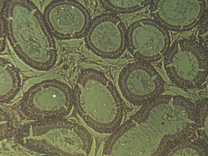
seminiferous tubules in the testis (left) and in the epididymis (right)
another cross-section of seminiferous tubules in the testis
In females, the Wolffian duct degenerates (apoptosis??) I don't know!
In vertebrates, the same piece of tissue (genital ridge) differentiates into either testis or ovary.
Primordial germ cells aggregate there, guided by an unknown mechanism.
The primordial germ cells appear brown in this photograph.
Myotomes right under them
Then sclerotomes
The continuous blue stripe on the right is the neural tube.


(female sex ducts develop from lateral plate mesoderm)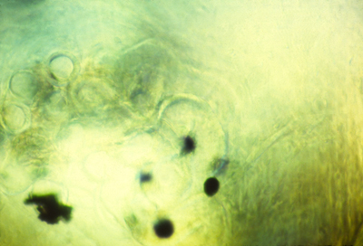
The pronephric ducts connect to the cloaca, at the rear.
(The ureters form from buds off the base of the pronephric duct,
extend forward, and induce differentiation of the metanephros.
Urine from it is carried to the cloaca through the pronephric duct.
Just to make things harder, the "Wolffian Duct" is a synonym for the pronephric duct, & sometimes people call it the mesonephric duct!
and it begins with a very simple geometric structure,
which becomes more and more complicated,
dividing into four chambers,
while beating and pumping blood the whole time!
while the motor is running full speed the whole time!
(by "cavitation" + by mesenchymal cells changing into two epithelial sheets, with their apical surfaces facing each other, and fluid between them
The splanchnic layer
in contact with the endoderm below it.
The somatic layer
in contact with the somatic ectoderm above it.
The space between these two layers is the coelomic cavity (=coelom),
This becomes filled with coelomic fluid, which is secreted into it by the surrounding cells.
the extreme right and left sides of the coelomic cavity becomes continuous with the space between the chorion and the amnion,
a fluid-filled space which is called the extraembryonic coelom.
& forms where the right & left lateral plate mesoderm come together, slightly anterior to the neck. (is its original location
Alternative possible experimental results!
1) No heart develops, on either side?
If pieces of plastic or other physical barriers were inserted where the heart normally forms, so as to block the right and left sheets of lateral plate mesoderm from coming together !!????
2) "Half-hearts" develop: incomplete structures on each side?
3) A heart develops only on the left side?
4) A heart develops only on the right side?
5) Two complete, normal hearts develop (one on each side)?
(one heart on each side of the barrier)
then each half can develop into a whole leg, or wing, arm, etc.
(whose muscle and bone structures are mirror images of each other)
The heart beats even before the kidneys start functioning.
Mesonephros the tubules of which connect to the Wolffian Duct
Metanephros has an entirely new and separate duct = the ureter
If you donate a kidney, check first to make sure you have 2.
Mesonephric kidney tubules connect to seminiferous tubules!
The "vas deferens"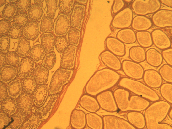
A separate "Müllerian duct" develops from lateral plate mesoderm
(in both sexes!)
But then the Müllerian duct degenerates in males.
The Müllerian duct becomes oviduct, fallopian tubules, & uterus.
These cells do NOT derive from ectoderm, mesoderm or endoderm.