Micrographs of tissue culture cells
These are photographs taken in previous years of tissue cultures similar to those you prepared in the lab last week.
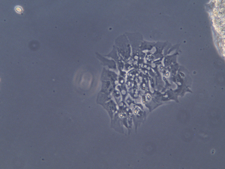
cells growing out of an explant
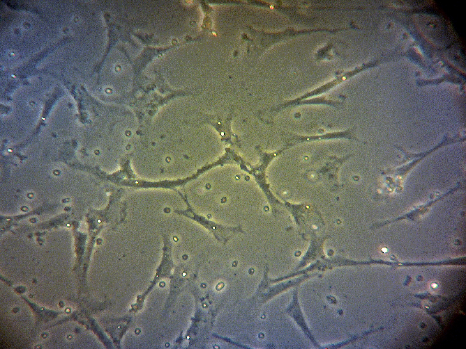
heart cells, taken through the microscope eyepiece
heart cells at higher magnification
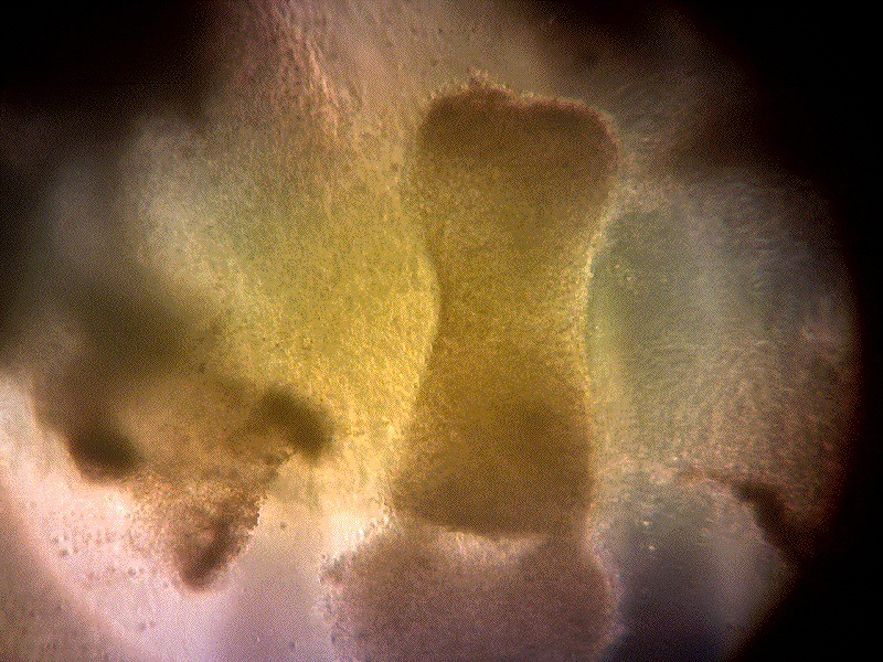
low-resolution photograph of leg cartilage, from which cells have migrated out
similar to the previous, but in this case the cartilage came from the eye
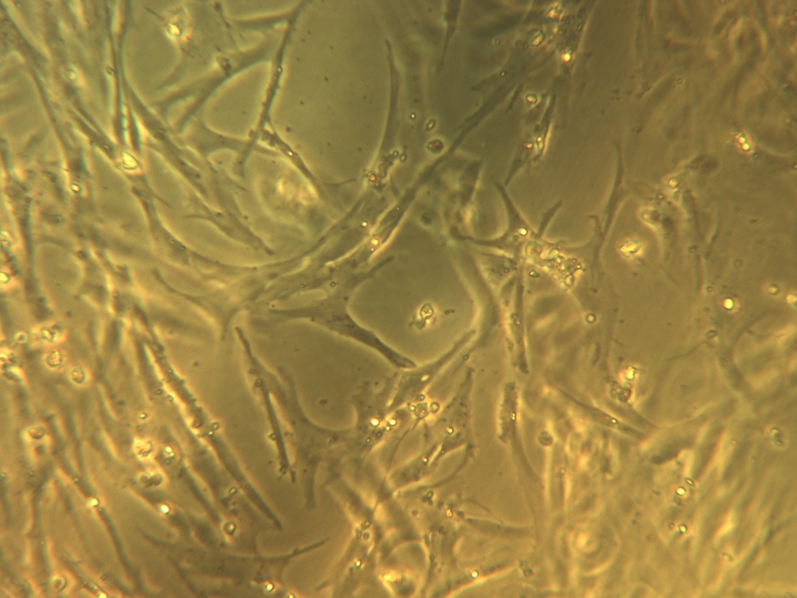
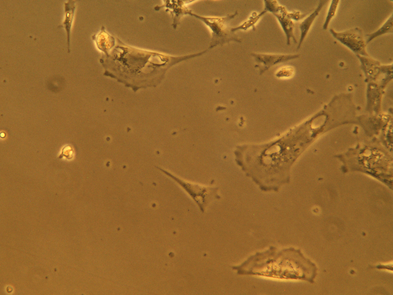
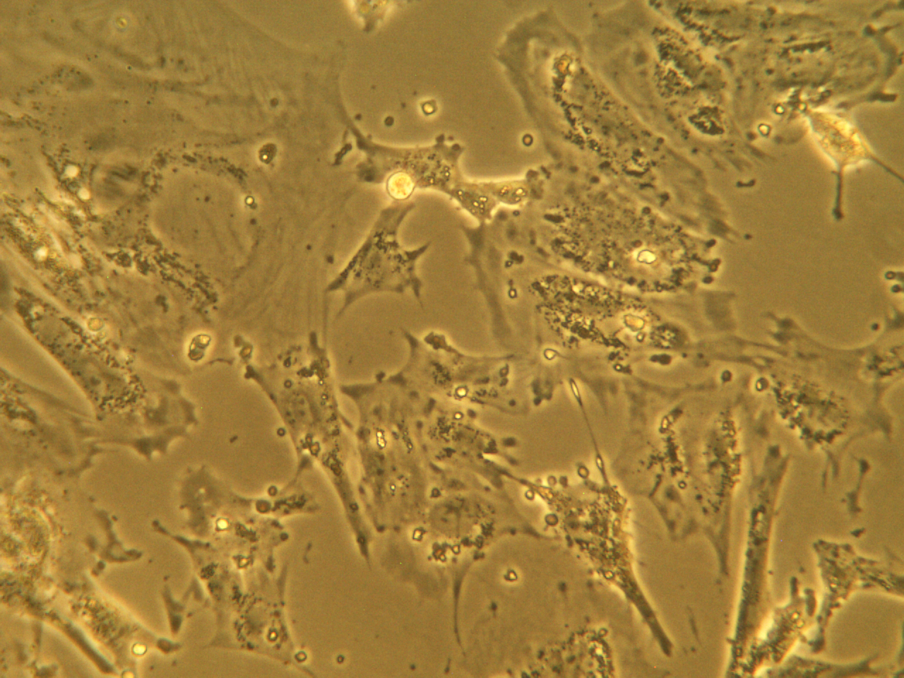
fibroblasts, typical of heart or leg muscle
another picture of fibroblasts
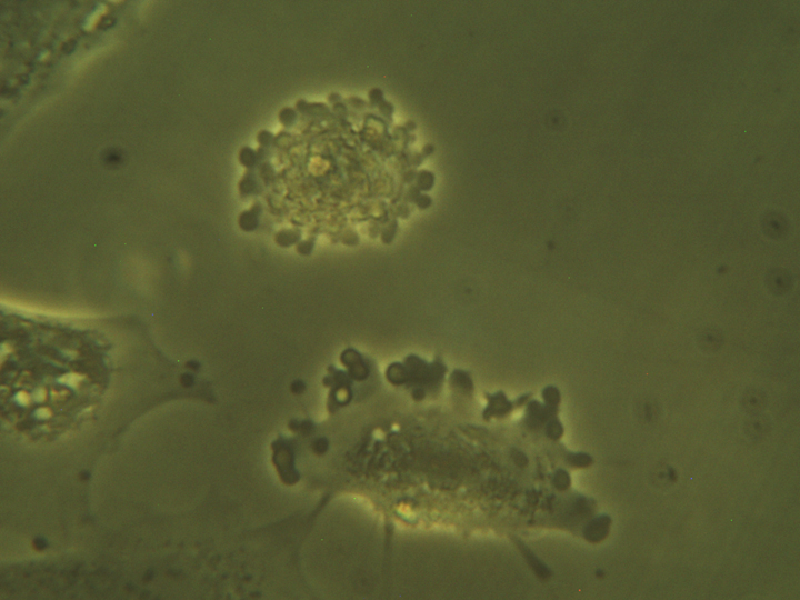
cluster of chondrocytes (above), glial cell (below)
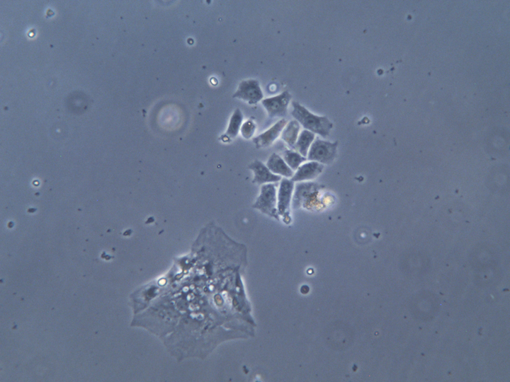
two cells resulting from mitosis, starting to spread out again
blebbing cells
chondrocytes at high magnification
island of cells, maybe nerves and myelin?
chondrocytes and fibroblasts from leg
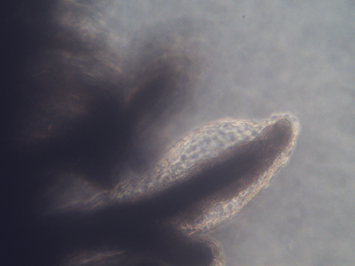
embryonic feather
The rest of the pictures are from the cultures you prepared last week.
explanted chunk of heart tissue
heart fibroblasts
two heart cells that have detached and are dividing
liver cells
liver cells, higher magnification
liver cells; the cell in the center has rounded up, leaving retraction fibers attached to the substratum
more liver cells
cells growing out from a piece of pigmented retina
retina; note the black pigment in the center
myotubes in a culture made from cartilage
Please make drawings of at least five different cell types, as if you were looking at your cultures in the lab. Please e-mail your completed sheets to your TA by April 24/25.
| 