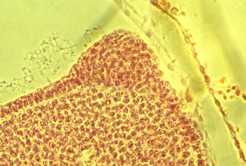
The Apical Ectodermal Ridge ("AER") is a long narrow thickened stripe of the outer layer of skin running down both sides of all vertebrate embryos (except salamanders!), at the outer tips of legs, wings, arms and fins.

Surgical removal of the AER (and also genetic inhibition) causes distal parts of legs not to form.
The sooner the AER is removed, the more proximal parts of the legs fail to develop.
For example, if the AER is removed later, then just the foot fails to develop. If removed earlier, then no wrist.
Before continuing, let's invent possible explanatory theories:
What are explanations? Imaginary "facts" that correctly predict
Especially if they make more predictions than assumptions. That suggest causal connections between sets of facts that previously had seemed unrelated to one another.
I) Maybe the AER secretes proteins that diffuse to form a diffusion gradient in the proximo-distal axis.
(This was disproven; Can you figure out how.)
II) Maybe each AER secretes a series of different stimulatory proteins?
First it secretes chemicals that induce formation of shoulder, etc. Those proteins that induce hand get secreted last... etc.
(this was also disproven; let's figure out how.)
III) Maybe the length of time inner tissues are close to the AER controls proximo-distal skeleton condensation.
Short time of being close to AER causes humerus, longer time radius & ulna.
(This was long taught as fact, but is now unpopular; lets figure out how to test it)
IV) Maybe shoulder stimulates humerus condensation, then humerus stimulates condensation of radius & ulna, and they stimulate formation of carpals, etc. but these sequential inductions can't occur unless the AER sends some signal to the condensing cells?
Let's invent experimental tests of this category of theory.
A scientific test is a situation in which alternative theories make different predictions. Preferably opposite predictions.
If you surgically cut off the AER, but implant a plastic bead that contain Fibroblast Growth Factor, then normal skeletal development occurs (despite the absence of the AER. Please invent theories that predict this.
A certain mutation in chickens caused two wings to develop on each side, analogous to a biplane.
Surgical grafting of a second AER above or below the normal one results in formation of two wings, one above the other (again, sort of like a biplane).
Please suggest interpretations of these experimental observations.
What hypotheses do they support, for example which theories about signals from the AER.
Suggest possible reasons why the AER cells need to be different elongated shapes in order to accomplish their function of signaling (pinched together at the apical ends, in birds & fish; and pinched at both ends in mammals)
In no kind of animal are AERs more than one cell thick, as you might guess on the basis of them being "thickenings".
The AER exists only during embryonic development, then its cells become the same as other cells of the outer layer of the skin (in the area of your finger-tips.
In fish and tadpoles, very similar thickening forms along the back and tail of fish, and develops into fins. This was part of the evidence supporting the "Fin-Fold Theory" (of the late 1800s), that arms, legs, wings, and fins all evolved from several very long continuous fins that originally extended from head to tail, top and bottom, and along both sides.
Please invent a molecular version of such a fin-fold theory . (Some of the same signaling proteins are concentrated in tail fins as are also concentrated in the AER of arms and legs).
If you take a plastic bead soaked in fibroblast growth factor, and implant it along the side of a salamander embryo, between the front and hind limb buds, it will stimulate formation of a fifth leg. (or a 5th & sixth leg, if you put implants on both sides)
The original discovery of 5th leg induction was by Balinsky, who implanted ear and nose tissue, instead of FGF-coated beads. Please invent possible connections of 5th leg induction and the fin fold theory.
Please try to invent theories that can explain (= correctly predict, = make sense of, = lead to some additional)
why salamanders are the only vertebrates that don't have AERs, and also the only vertebrates that can regenerate legs.
In other words, please invent possible connections between being able to regenerate legs and not forming AERs.
* It's just a coincidence...?
** Maybe salamander skin forms the same signaling proteins as the AER, except for some reason the cells that secrete these molecules in salamanders don't have any special thickened or pinched-together shapes?
*** Maybe if an AER could be caused to form on the stumps of amputated mammal or bird legs or wings, then they would become able to regenerate...?
**** Maybe the ability to form 5th legs along the sides depends on not having (and not needing) an AER.
Incidentally, during regeneration, salamander legs DO form a thickening of the outer layer of the skin at the tip of the bud.
(This thickening is not elongated in the anterior-posterior axis, however, and is not called an AER.)
What would you expect to happen if you surgically removed this thickening (hint: at different times...?)
What if you transplanted this thickening to an abnormal location...?
What if you could stimulate the outer layer of the skin to thicken? In mammals & birds? Between front & hind legs?
In some recent papers, the AER has been called "a timing device". Would you agree? In what sense?
The proximo-distal axis of salamander legs has a gradient of whatever property of cells causes sorting out of dissociated and randomly mixed cells. Specifically, "hand" cells sort out to interior locations relative to wrist, wrist interior to elbow, elbow interior to shoulder; likewise hand gets engulfed by...= invaginates into wrist cells; wrist invaginates into elbow cells.
For scientists who believe in Steinberg's Differential Adhesion Hypothesis, engulfment, sorting out and invagination are caused by stronger cell-cell adhesiveness of whichever cells get engulfed, invaginate into the others, and sort out to the more interior location. Therefore, the DAH concludes = predicts a gradient of stronger cell-cell adhesiveness from shoulder (weakest adhesion) to foot (strongest adhesion). The other theory of cell sorting says that interior sorting / invaginating into = getting engulfed results from stronger cell contraction along surfaces of cell aggregates and interfaces between cells of different organs. So distal limb cells should contract more strongly.
Another not-yet-explained set of discoveries is that if elbow-level cells are grafted to the foot (in a salamander larva), then the grafted mass of cells moves up the leg, back to the elbow area, where they came from. The reverse also happens: Hand level cells that are grafted to the elbow get pulled or pushed (or whatever) back to the hand area. Grafted tissues move back to whatever level of the proximo-distal level that they were originally located. Supposedly they are pulled by maximization of cell-cell adhesion.
Question: Would this really work? Could these migrations be explained (predicted) by a proximo-distal gradient of cell contractile strength along boundaries (interfaces) between elbow cells and wrist cells, etc.
Note: Don't assume that these differences in contractile strengths (or differences in forces exerted by adhesions) would not need to exist all the time. Consider how the predictions differ if contact between elbow cells and wrist cells were to stimulate development of forces in the interface where they contract each other. Notice that for grafted tissues to get pulled in a particular direction, what is needed is stronger contraction (or more adhesion) on the side of the graft toward which the grafted cells are pulled.
BODYShoulderUpperarmElbowForearmWristHand
BODYShoulderUpperarmElbowForearmWristHand
Humerus Radius & Ulna Carpals Digits
Wrist graft---> Stronger limb here? But why stronger here??? <---Elbow graft
BODYShoulderUpperarmElbowForearmWristHand
BODYShoulderUpperarmElbowForearmWristHand
<-same Wrist graft same-> same strength Elbow graft same strength
BODYShoulderUpperarmElbowForearmWristHand
BODYShoulderUpperarmElbowForearmWristHand
Wrist graft---> WHY stronger here??? WHY stronger here??? <---Elbow graft
BODYShoulderUpperarmElbowForearmWristHand
BODYShoulderUpperarmElbowForearmWristHand
because limb tissue is stronger? because graft is stronger?
Elbow graft --> why? ???<--- Elbow graft
BODYShoulderUpperarmElbowForearmWristHand
Do grafted tissues change strength?
A good paper about the AER:
Fernandez-Teran, M. I. and Ros, M. A. (2008). The Apical Ectodermal Ridge: morphological aspects and signaling pathways. Int J Dev Biol. 52(7):857-71
Departamento de Anatomía y Biología Celular, Universidad de Cantabria, Santander, Spain.
Abstract
The Apical Ectodermal Ridge (AER) is one of the main signaling centers during limb development. It controls outgrowth and patterning in the proximo-distal axis. In the last few years a considerable amount of new data regarding the cellular and molecular mechanisms underlying AER function and structure has been obtained. In this review, we describe and discuss current knowledge of the regulatory networks which control the induction, maturation and regression of the AER, as well as the link between dorso-ventral patterning and the formation and position of the AER. Our aim is to integrate both recent and old knowledge to produce a wider picture of the AER which enhances our understanding of this relevant structure.
PMID: 18956316 free full text
Benazet, J. D. I, and Zellare, R. (2009). Vertebrate limb development: moving from classical morphogen gradients to an integrated 4-dimensional patterning system. Cold Spring Harbor Perspectives on Biology. 2009 Oct;1(4):a001339. doi: 10.1101/cshperspect.a001339.
Abstract
A wealth of classical embryological manipulation experiments taking mainly advantage of the chicken limb buds identified the apical ectodermal ridge (AER) and the zone of polarizing activity (ZPA) as the respective ectodermal and mesenchymal key signaling centers coordinating proximodistal (PD) and anteroposterior (AP) limb axis development. These experiments inspired Wolpert's French flag model, which is a classic among morphogen gradient models. Subsequent molecular and genetic analysis in the mouse identified retinoic acid as proximal signal, and fibroblast growth factors (FGFs) and sonic hedgehog (SHH) as the essential instructive signals produced by AER and ZPA, respectively. Recent studies provide good evidence that progenitors are specified early with respect to their PD and AP fates and that morpho-regulatory signaling is also required for subsequent proliferative expansion of the specified progenitor pools. The determination of particular fates seems to occur rather late and depends on additional signals such as bone morphogenetic proteins (BMPs), which indicates that cells integrate signaling inputs over time and space. The coordinate regulation of PD and AP axis patterning is controlled by an epithelial-mesenchymal feedback signaling system, in which transcriptional regulation of the BMP antagonist Gremlin1 integrates inputs from the BMP, SHH, and FGF pathways. Vertebrate limb-bud development is controlled by a 4-dimensional (4D) patterning system integrating positive and negative regulatory feedback loops, rather than thresholds set by morphogen gradients.
Lu, P.L., Yu, Y., Perdue, Y., and Werb, Zena (2008). The apical ectodermal ridge is a timer for generating distal limb progenitors. Development. 135(8):1395-405.
Abstract
The apical ectodermal ridge (AER) is a transient embryonic structure essential for the induction, patterning and outgrowth of the vertebrate limb. However, the mechanism of AER function in limb skeletal patterning has remained unclear. In this study, we genetically ablated the AER by conditionally removing FGFR2 function and found that distal limb development failed in mutant mice. We showed that FGFR2 promotes survival of AER cells and interacts with Wnt/beta-catenin signaling during AER maintenance. Interestingly, cell proliferation and survival were not significantly reduced in the distal mesenchyme of mutant limb buds. We established Hoxa13 expression as an early marker of distal limb progenitors and discovered a dynamic morphogenetic process of distal limb development. We found that premature AER loss in mutant limb buds delayed generation of autopod progenitors, which in turn failed to reach a threshold number required to form a normal autopod. Taken together, we have uncovered a novel mechanism, whereby the AER regulates the number of autopod progenitors by determining the onset of their generation.