Slides shown on September 9:
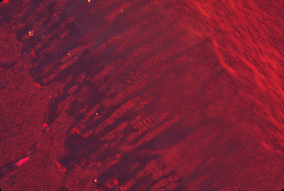
Both these slides show formation of the epiphysis:
Epiphyseal "growth zone" from a rabbit bone.
This is from the Wikipedia article on the epiphyseal plate
=======
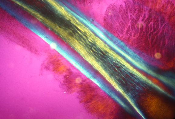
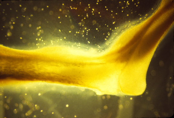
This image appears in Harris, Albert K. (2014). Connective Tissue Development, in
"Cell and Matrix Mechanics",
eds. R.R. Kaunas and A. Zemel, Taylor and Francis Group, Boca Raton FL.
UNC library link
The original caption is as follows:
Epiphysis of a rabbit bone, photographed by polarized light with quarter-wave filters, so that colors of birefringent materials will vary as a function of direction. Cartilage in the upper right area is gradually being destroyed and replaced by bone, advancing from the lower left, into the center. The yellow color results from collagen fibers in the cartilage being oriented transverse to the advancing bone. The dark blue streaks in the center show that collagen has become reoriented by 90°, so as to become parallel to the direction of the advancing bone. Stacks of enlarged chondrocytes are visible in the pale areas between the blue streaks.
Slides shown on September 14:
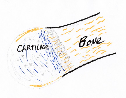
This is a sketch of a developing bone, similar to the epiphysis slides above.

Leg "bone" (at this stage entirely made of cartilage) seen by polarized light microscopy. Areas in which collagen fibers are predominantly longitudinal appear blue, while yellowish red indicates that collagen fibers are predominantly circumferential. The colors in this photograph indicate that there are many longitudinal fibers, but more circumferential fibers, perhaps 55% circumferential and 45% longitudinal. Living tissue. From Harris (2014), cited above
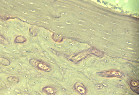
Ossifying bone photographed with polarized light
Section of a bone
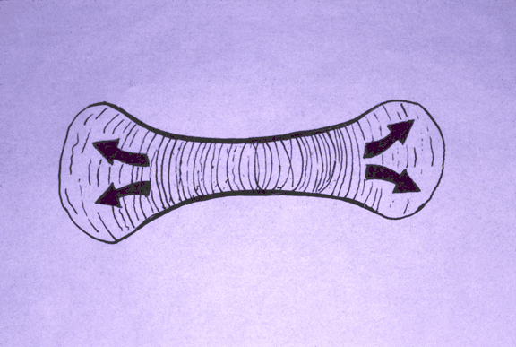
Drawing of forces in long bone shaping

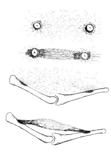
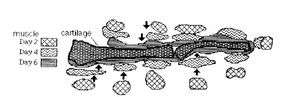
Photo montage of muscles aligned by cartilages
This figure is from Stopak and Harris (1982). Connective Tissue Morphogenesis by Fibroblast Traction. Developmental Biology 90, 383-398. Leg bones from 10-day old chick embryos were put in a culture dish with pieces of thigh muscle in a collagen gel. The collagen becomes aligned between the muscle explants and the bones as a result of cell traction, forming an anatomical pattern that looks like a natural leg.
A color slide of an artificial muscle formed in this way./i>
Drawings of theory of skeletal muscle formation by traction
Tracing of muscle explants pulled in by cartilages
Photograph of the head of a femur
Photograph of the head of a femur
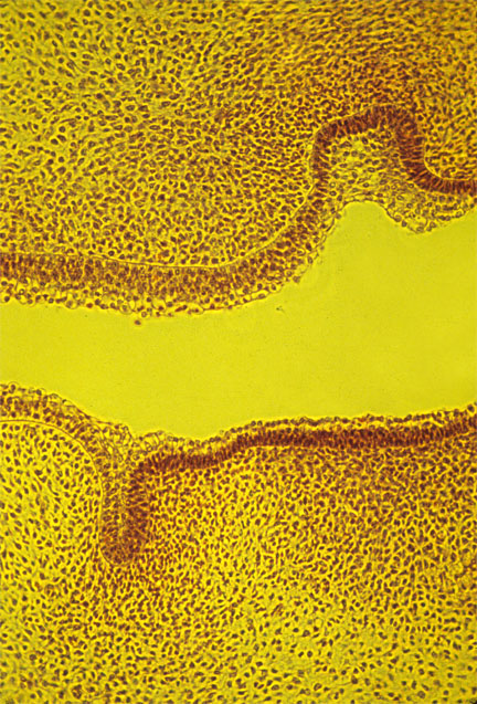
section of a molar
The dark area is empty space, where the enamel had been but broke off
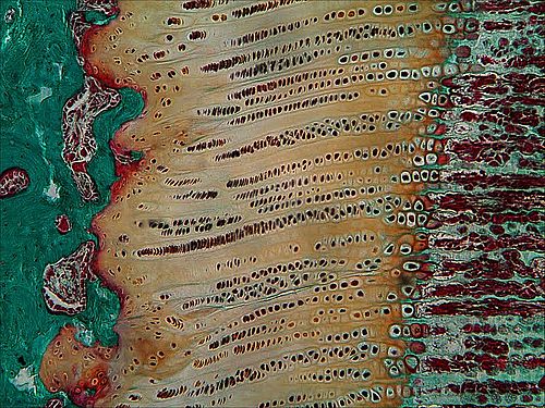
tooth rudiments
dentin and enamel
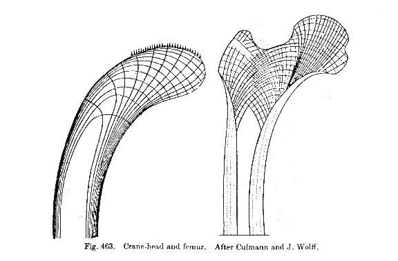 Drawing from the classic book On Growth and Form, by D'Arcy Thompson, showing the forces in the head of a femur bone.
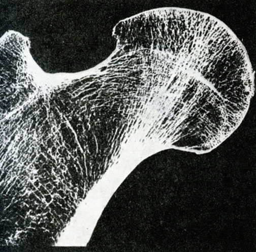
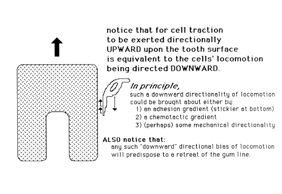
Tooth Formation
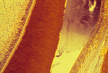
The outer layer of teeth (Ameloblasts --> Enamel) 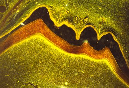
during preparation of the slide. The orange area is the dentin, which is a kind of bone.