Cells in tissue culture spread on the substratum and attach by "focal adhesions."
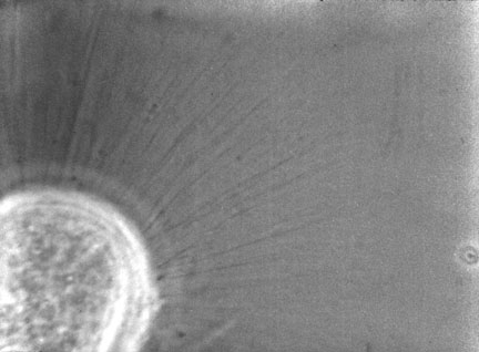
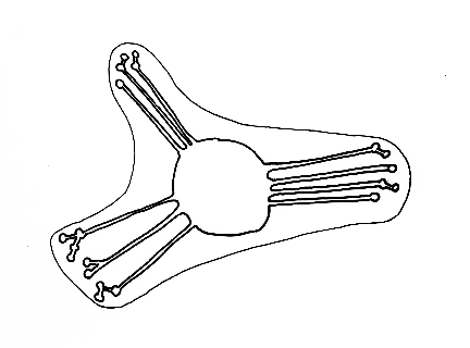
When they round up prior to mitosis, these adhesions persist, but the cell body draws away from them, leaving then "retraction fibers." Sometimes these fibers break, leaving a blob of cytoplasm where the adhesion was. In most cases, however, after cytokinesis each daughter cell spreads out again, refilling the retraction fibers.
Look at the cell approximately in the center of the frame at about 0.04, and watch as it rounds up and divides (0.05-0.06).
Harris, A. K. (1973) Location of cellular adhesions to solid substrata. Dev. Biol. 35: 97-114.
doi: 10.1016/0012-1606(73)90009-2. PMID: 4787750
Two diagrams of what's happening when cells round up in mitosis:


video of cell undergoing mitosis and cytokinesis
Look for the retraction fibers.

photograph of a cell with many retraction fibers

Diagram of cell body with retraction fibers and their adhesion points
Retraction fibers have been mistakenly assumed to be actin filaments (filopodia) that extend outward from cells:
Paper by Vasioukhin et al. (2000).
A figure from this paper was shown in class.
Scanning electron micrograph of a HeLa cell, showing extensive retraction fibers. [From Wikipedia]
Video of chicken cells cultured on silicone rubber.
When EDTA is added to the culture (about 20 seconds into the video), the cells begin to detach from each other.
Compare this to blobs of mozzarella cheese detaching
Digiorno Four-Cheese Rising Crust Pizza topped with some delicious Italian sausage from Coon Rock Farm, Hillsborough NC