How cell differentiation is controlled.
The signals that cause each cell type to differentiate at the correct places.
The mechanical forces by which cells move each other from place to place.
It includes the anatomy of embryos, cell rearrangements, gene activation, cell movements, a little about the physics of cells, concepts of symmetry, small amounts of computer simulation, regeneration, aging, cancer etc.
How do bones get their shapes?
What connects muscles to bones?
What guides nerves to their correct connections?
What forces shape the eye, the lens and the cornea?
What goes wrong that allows cancer cells to invade?
How can salamanders regenerate legs, but people can't?
+++++++++++++++++++++++++++++++++++++++++++++++++++++++++
Regarding this year's textbook:
Previous years, I have often used Scott Gilbert's excellent "Developmental Biology"
(which I still recommend as a good source book)
[Although it contains very little on mechanical forces and mathematical modeling
and what it does say is on those subjects is completely wrong, without exception.]
Partly because of the high price ($ 150.00+), of Gilbert's book, and because I think it has lost its focus and sharpness, this year we will experiment with Jonathan Slack's "From Egg To Embryo: Regional Specification in Early Development"
(paper-back of the second edition is about forty dollars)
(used copies may be cheaper)
The two main elements of embryology are
I) Signaling, to control where cells will differentiate into each cell type.
II) Mechanical forces that move and shape cells, and cell secretions.
Of these two areas, Slack's book concentrates on the former
My own research concentrated on the latter. Therefore, I can provide original web page material.
Except for cost, I would have also have adopted a second book: Jonathan Bard's "Morphogenesis: The cellular and molecular processes of developmental anatomy".
which is a companion book to Slack's, in the same series.
////////////////////////////////////////////////////////////////////////////////////////////////////////////////////
1) Differentiated cell types (about 250 cell types in humans; Slack's book says somewhere between 200 to 300)
10 specific examples:
- red blood cells
pigment cells of the skin
pigmented retina cells
the blue-sensitive cone cells of the retina
the green-sensitive cone cells of the retina
the red-sensitive cone cells of the retina
ganglion cells (that form the optic nerve) [but are there 3 kinds?]
macrophages
skeletal muscle cells
cardiac muscle cells
liver cells
Oogenesis and spermatogenesis are examples of cell differentiation:
Egg and sperm precursor cells migrate from a distant location. Their differentiation is determined by special cytoplasms.
2) Oogenesis (=development of oocytes = egg cells)
-
Enlargement,
Storage of yolk,
Over-activity of genes,
Swelling of nucleus,
bird egg as compared with mammal or sea urchin egg.
Even mammal egg cell becomes 1000 times size of ordinary cell;
bird eggs are millions of times bigger than a normal cell.
Meiosis is delayed
in most species (including humans) meiosis is not completed until after fertilization.
Sea urchin eggs are unusual in already being haploid when fertilized.
Dog oocytes undergo both meiotic divisions after fertilization.
Oocytes need all 4 sets of chromosomes for as long as possible,
To support their very high rates of RNA synthesis & storage.
3) Spermatogenesis:
-
differentiating sperm become smaller,
discard most of cytoplasm,
develop one, or in some species 2, or more, flagella,
Nucleus becomes smaller, inactive, less transcription
Both meiotic divisions occur before sperm cell differentiation.
4) Fertilization: Fusion of plasma membranes of sperm and egg cell; entry of sperm nucleus into the cytoplasm of the oocyte.
5) Mechanisms to prevent any more sperm from getting in
After one enters "polyspermy" is fatal for later development.
An oocyte has the equivalent of the resting potential;
trans-membrane voltage ~70 millivolts positive inside
Fertilization initiates the equivalent of the action potential;
except slower, and longer-lasting.
Sodium ion channels open,
positive sodium and calcium ions diffuse inward.
Cell membrane becomes positive inside,
which inhibits fusion of any more sperm
6) Cleavage: first dozen or so mitotic divisions
in many species rapid & synchronous,
in mammals slow and not synchronous.
In some species hindered by yolk; meroblastic cleavage.
In some species very consistent, nematodes being extreme example.
In other species not consistent (mammals being an extreme example;
and some wasps also having extreme variation)
[this is not just a lower-animal versus higher-animal difference]
7) Blastula stage blastoderm (in birds) blastocyst (in mammals)
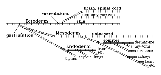
8) Gastrulation
subdivision of the developing embryo into 3 primary germ layers
ectoderm nervous system & outer layer of the skin (and also the lining of
the mouth)
mesoderm muscles, kidneys, heart, skeleton, and the inner layer of the skin
endoderm intestine, liver, pancreas, stomach, esophagus, lungs etc.

9) Neurulation Ectoderm subdivides into
-
Neural tube brain, neural tube, retina of eye, some other things.
Neural crest pigment cells of skin, sensory nerves, autonomic nerves, etc.
Somatic ectoderm: outer layer of skin, lenses of eyes, inner ear etc.
-
Notochord
Somites
Intermediate mesoderm
Lateral Plate mesoderm
-
Sclerotome (the cells of which will differentiate into skeleton)
Myotome (whose cells will differentiate into skeletal muscles ) Dermatome (the cells of which will become the inner layer of skin)
The Lateral Plate Mesoderm subdivides into... Coelom, Heart, etc.
Do you notice any patterns, in these different subdivisions?
Sample questions that might be on an exam, based on materials covered so far: (some of the ones toward the end involve some imagination and creativity)
a) Name at least 8 examples of differentiated cells in humans?
b) Meiosis occurs when, relative to times of fertilization and cell differentiation?
(hint: the answer is very different in oogenesis as compared with spermatogenesis)
c) Why are most mammal embryos triploid for the first 2 or 3 hours after fertilization?
(And are any kinds of mammals pentaploid for a while after fertilization)
d) What is an example of a kind of animal whose oocytes are haploid before fertilization?
(and if you can't think of any, then cniderians are the only other example)
e) Do unfertilized oocytes have a voltage difference between cytoplasm as compared with surrounding fluid?
f) What voltage change occurs at fertilization?
g) How similar to, but how different than, the conduction of a nerve impulse?
h) What purpose could be accomplished by artificially stimulating this voltage change?
i) Could the same purpose be accomplished by a drug that inhibited oocytes from undergoing this voltage change?
j) What do embryologists mean by "the cleavage stages" of development?
k) What happens during gastrulation.
l) What are the names of the three primary germ layers?
m) When embryology books use the word "germ" (as in "germ layers") does it have anything to do with disease-causing bacteria or viruses? Hint: no
n) From which germ layer does the nervous system develop?
o) Muscles, bones, kidneys (& what else) develop from which embryonic germ layer?
p) Lungs, liver, pancreas, (& what else) develop from which germ layer?
q) The outer layer of the skin develops from what?
r) What about the inner layer of the skin? What does it develop from?
s) The 3 germ layers form during the process of what?
t) During neurulation, what becomes subdivided into which three parts?
u) What is meant by polyspermy? Why is it bad?
v) Why would Darwinian selection NOT favor mutant organisms in which the sperm were more able to penetrate into oocytes that had already been fertilized by another sperm?
w) Would the answer to the previous question be different if this mutation also caused sperm to be able to destroy or otherwise remove the sperm that had already fertilized the oocyte? (Hint: Yes; but figure out and explain why?)
x) Which aspect of actual embryonic development is most surprising, or different from what a sensible person (you) would have guessed?
y) Imagine you are writing a science fiction book, and inventing stages of embryonic development for the animals on another planet; Invent sequences of events that are as different as possible, at every stage, than what occurs in animal development on earth.
z) Guess what might be the molecular-genetic basis of the primary germ layers? What might be their evolutionary cause?
Hint: Huxley used the words endoderm and ectoderm to refer to the inner and outer epithelia of Hydra. Was he implying any particular hypothesis about embryos?
Can you suggest a hypothesis about relations between endoderm in adult Hydras and in embryonic vertebrates?