Lecture notes for Friday, January 23, 2015
Changes in strengths and directions of forces cause changes in shapes of cells, embryos and organs
Increased curvature being caused by decreased tension in the cortex of a fertilized teleost oocyte:
Continue with a digression about mammal polar bodies & how big they are.
video: mammal egg & polar body
video: Mammal egg being fertilized
Are all cell cleavages caused by localized inhibition of contractile tension of the cell cortex?
Answer: Scientists who specialize in mitosis have debated for many years whether cell cleavage furrows are formed by signals that strengthen cortical contractility at the "equator" (half way between the poles of mitotic spindles) versus whether cleavage furrows are induced by signals that weaken cortical contractility near the poles of the mitotic spindle.
Although I have been on the side of stimulation in that debate, these videos of blastodisk formation in fish embryos and polar body formation in mammal embryos give more support to the theory that the cell cortex is weakened in circular areas around the animal pole!
What do you thing about this? What experiments should be done?
Later in the course you will learn about other examples of embryonic structures being re-shaped by changes in cortical contractility (= contractile tension)
Tension often changes in amount, and sometimes changes in direction.
For example, look again at the Fundulus video
and notice the first cleavage division.
Does the curvature of the cell surface increase or decrease?
(in the cleavage furrow? At other parts of the cell surface?)
Notice small amounts of cytoplasmic flow in the cell cortex. Sometimes this is toward the animal pole; other times the flow is away from the animal pole.
Please invent a hypothesis that can explain what you see. What predictions does your theory make, for example about the next divisions, from 2 to 4 cells and from 4 to 8 cells?
Suppose that you had a chemical that could inhibit cell cortical contractility: invenr some experiments that you could do with it.
During each cleavage, both sides in the debate about weakening or strengthening the cell cortex agree that tension has to become directionally stronger in the direction along the cleavage furrow, and/or become weaker in the direction transverse to the
cleavage furrow.
On an exam, could you draw a map of the locations where tensions change in the cell surface (cortex)?
Could you draw a graph of changes in tension as a function of time?
What about locations of changes in cytoplasmic flow, and of cytoplasmic pressure?
Consider the shape of an eyeball: spherical. with one bulge (the cornea).
On an exam, could you argue pro or con whether the shape of eyeballs and corneas are caused by amounts of tension in the surface of the eyeball and the cornea?
Is genetic control of tension possible?
Is there any other way for genes to cause the shapes of the eyeball and the cornea.
Do you know what "Lasik Surgery" means?
If you needed to change the curvature of someone's cornea, would you rather do it by cutting away parts of the cornea, or by weakening or stengthening tension in the cortex?
If you invented a drug or other treatment that could cause changes in the tension and/or the flexibility of the cornea, how could your treatment help the near-sighted become far-sighted or the reverse?
What about if your treatment could change the tension in the surface of the lens?
Human eyeballs have a safety-valve that lets fluid leak out of the eyeball, into the blood. It is called "The Canal Of Schlemm." This canal is located at the outer edge of the cornea, right where the curvature changes. (extending 360 degrees all around the edge of the cornea)
See image from Schlemm's canal article on Wikipedia
Imagine a graph of amount of fluid outflow as a function of differences in the tension of the eyeball (and the fluid pressure inside the eyeball) Sketch a shape for this graph that would cause the fluid pressure to change by the least amount if there were to be an increase in the amount of fluid being secreted into the interior of the eye. The group of diseases called "Glaucoma" involves increased fluid pressure in the eye and progressively causing blindness.
My point is that medically important phenomena often involve effects of forces on anatomical shapes
------------------------------------------------------------------------------------------------
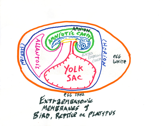
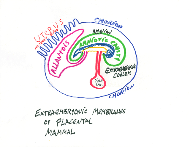
Gastrulation in Birds, Reptiles and Mammals
Mammal gastrulation (including human gastrulation) is very similar to what you see in this video.
The following time lapse video shows gastrulation and neurulation of a very early chicken embryo. The head end is toward the right and the posterior is toward the left.
Please watch this video carefully and try to figure out the locations and directions of the different pulling and pushing forces that rearrange tissues and create the body axis. (the visible time clock is not accurate, and greatly understates the duration of these events.)
Closure of the neural tube occurs gradually from right to left.
Future mesoderm cells are still ingressing down away from the upper surface, with the future notochord cells (barely!) visible as a dark point called Hensen's node (=the primitive node)
At any location along the embryo's anterior-posterior axis, the last future mesoderm cells to ingress become the notochord. Future notochord cells are internalized at the Hensen's node (better to think of the internalization of future notochord cells as creating a small, barely visible dark dot) At the start of this video, Hensen's node is just to the right of the exact center of the field of view. It slowly moves toward the left (toward the posterior).
It may help to imagine Hensen's node as analogous to a zipper, closing the process of ingression of future mesoderm and endoderm.
This rearward movement is called "The Regression Of The Node".
It is how we gastrulate. The Hensen's node is equivalent to the dorsal lip of the blastopore, in two important ways: #1) Its cells will differentiate into the notochord. #2) If you graft node cells to another part of the embryo, they will induce a second neural tube and body. Conrad Waddington discovered this.
--------------------------------------------------------------------------------------


