Lecture notes for Wednesday, January 24, 2018
Any unicellular organism that changes shape as it moves (and isn't ciliated) most biologists will regard as "amoebae". Its as if cars and airplanes, trains, ships and rockets were regarded as using the same method of propulsion. Cancer researchers sometimes study amoebae, on the assumption they are basically the same; and maybe they are not completely wrong.
First Key Fact: There are about a dozen different kinds of amoeboid locomotion, fundamentally different from each other. The total number isn't known.
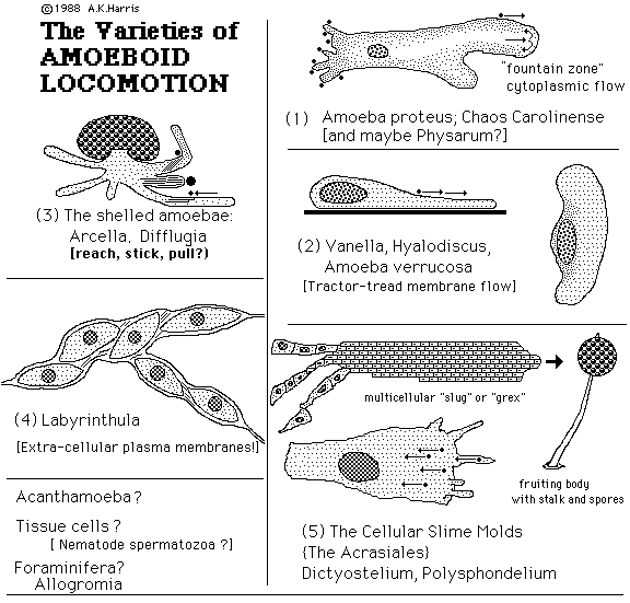
Specific Examples of different kinds of amoeboid locomotion:
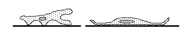
I) "Labyrinthula" Has several layers of plasma membranes outside the cells, with extracellular cytoplasm "slimeways".
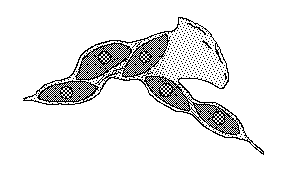
99% of biologists don't know that labyrinthula exist, or anything like them.
Labyrinthula are very common in the ocean, and cause diseases in sea grasses, and perhaps other organisms. Fresh water and land species also exist. Some of the latter attack golf courses!
There is a Wikipedia article about Labyrinthula, which in my opinion isn't very good, has no illustrations, but does have a good bibliography. David Porter, at the U of Georgia has done the best research on these interesting creatures.
Excellent YouTube video of Labyrinthula: https://www.youtube.com/watch?v=yFK8D41VvLY
II) Rolling Amoebae (a name that I just invented, for lack of anything better.)

Their plasma membrane flows forward across the top, and rearward on the bottom (or along whatever surface they get attached to).
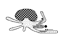
III) Shelled amoebae. These secrete and/or build hard little shells, sort of like microscopic snail shells. These really do reach out long narrow cytoplasmic protrusions, which stick to anything, and contract, pulling the cell forward, and pulling smaller objects rearward.
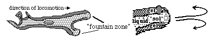
IV) The kinds of amoebae that are used so much for teaching introductory biology courses: Amoeba proteus and Chaos carolinensis. (the latter discovered by a UNC Biology professor, H.V.Wilson. Go Heels!)
 :
: 
These have rapid cytoplasmic flow. In fact, most research papers about them are really attempts to understand how this flow works. But it is not so clear how their cytoplasmic flow causes movement of the cells (although it definitely does!) The "gelling" of amoeba cytoplasm is caused by polymerization of cytoplasmic actin
video of Amoeba proteus moving, showing cytoplasmic flow
video of an amoeba engulfing some food
Forward transport of marker particles attached to the outer surface of the plasma membrane;
And rearward transport of attached particles where pseudopodia are retracting.
V) Amoebae that transport attached particles rearward.
Dictyostelium and other cellular slime molds.
Dictyostelium cells move as individuals, phagocytizing bacteria, and then as multicellular "slugs". This aggregation is induced to occur when cells "attract" each other by chemotaxis; the attractant substance (for D. discoideum) is cyclic AMP.
video of Dictyostelium amoebae
White blood cells, nerve cells, mesenchymal cells, etc, etc, etc. move in a similar way.
All component cells of multicellular animal bodies.
A shared property of this class of amoeboid locomotion is that if particles, antibodies or most other markers are attached to the surface of their plasma membranes the result is always active directional transport in the direction that traction is being exerted. This is called "retrograde surface transport".
Almost certainly it results from traction being applied to small, moveable objects. Actin molecules polymerize into fibers along the front-most parts of cells, and these fibers get pulled rearward by active sliding along myosin fibers in more rearward and central parts of the cell.
Traction results from physical connections through the fluid mosaic structure of the plasma membranes. The locations where traction forces are exerted are several microns behind the leading edge. In some cells, filopodia are part of this exertion of traction.
Retrograde surface transport also occurs in Dictyostelium and other slime molds.
--------------------------------------------------------------------------------------------------
Osmotic swelling is a very important force in shaping embryos.
Cartilage expansion (which is not really "growth") is driven by a special version of osmosis called electroosmosis. This does not depend on semi-permeable membranes, like ordinary osmosis.
Electroosmosis is caused by positive ions (Sodium, potassium, hydrogen, calcium etc.) being held in gels by electric attraction toward negatively charged sulfate groups that are covalently attached to special chains of sugars (chondroitin sulfate, among others). The positive ions are trapped at high concentrations by this electrical attraction, which produces just as much pressure as would result from the same concentration of ions being held inside a semipermeable membrane. (22.4 atmospheres of pressure per one mole concentration of sulfates).
Vertebrate skeletons begin (mostly) as cartilages, much of which gets replaced by bones having the same shapes as the cartilage they replace. It is very important to understand that cartilage never changes into bone, although cartilages often get calcium salts deposited in them.
Except for the roof of the skull, the only way vertebrate embryos can make a bone that has any given shape is to secrete cartilage having that particular shape, and then replace the cartilage with bone.
If you squeeze a piece of cartilage, positive ions get pulled along with water that gets pushed out of cartilage. This creates a brief positive electrical charge, that can be hundreds of volts! (but very low amperage) Then you get a brief voltage in the opposite direction.
Some researchers think these voltages serve some function (rather than just being side effects of the use of electrical attraction as a way to exert and resist pressure). The suspected function is stimulation of bone production. It has definitely been proven that bones get stronger whenever and wherever more forces are exerted on them. For example, tennis players develop much stronger bones in their right arm if they are right-handed.
At a research meeting I went to a couple years ago, people reported that stressed skeletons produced voltages by piezoelectricity, which is a different phenomenon that occurs in many crystals.
Medically, there is an enormous need to discover methods that can produce strengthening of bones. Electric currents are one of the methods that has been tried.
------------------------------------------------------------------------------------------------
Expansion of fluid-filled spaces. The brain is a hollow sac, inflated with salty water.
Lobes of the brain result from mechanical strength being made weaker in certain places.
Reducing the pressure will cause abnormal brain development.
The eyeballs also develop into fluid-filled balloon-like structures, which have been shown to develop abnormally if punctured so that the fluid leaks out. The inner ears also go through a stage of being balloon-like, in a mechanical sense. There may be other examples of hollow embryonic structures that develop by fluid pressure, but I can't think of any.
------------------------------------------------------------------------------------------------
Fluid inflation in relation to development of teleost fish embryos.
Teleost fish form their brains and spinal cords by hollowing-out a solid rod of cells.
Almost all other vertebrates form their brains and spinal cords by folding a flat sheet of cells and then fusing their edges together.
What do you think this difference might mean?
#1) That fish use fundamentally different mechanisms than other vertebrates to form their central nervous systems?
#2) That folding and hollowing out can both be caused by the same basic cell properties?
#3) Something else?