Lecture notes for Friday, January 26, 2018
Embryology of Teleost Fish
Zebrafish survive and go through their whole life cycle in captivity (and are not hard to feed)(Unlike, for example, the teleosts killifish and salmon, on which most research on fish development used to be done).
They are reasonably small, but not too small, not microscopic.
Zebrafish have a reasonably short life cycle (months, instead of years).
They lay eggs externally, in contrast to "live bearing species" in which development occurs inside the mother's body). Many dozens of eggs are laid per female.
The eggs and embryos are transparent.
They are not polyploid. (Xenopus laevis turned out to be tetraploid!)
Many mutant lines have been found and are being kept in "stock centers".
The genome has been sequenced, except for heterochromatin. ).
***********************************************************************
Teleosts have meroblastic cleavage.
Teleosts develop two extra-embryonic membranes:
-
The Enveloping Layer
The Yolk Syncytial Layer
The body of the fish develops entirely from the "Deep Cells"
These are crowded into a lens-shaped space below the hemispherical epithelial enveloping layer and the spherical mass of yolk below them.
During their gastrulation, the enveloping layer spreads over the yolk, surrounding it. This spreading is the fish's special way of gastrulating, and is called epiboly.
Deep cells crawl toward what will be the head and body axis, converging from both right and left sides.
By pushing down on the deep cells, you can easily induce formation of more than one head, and two or more body axes, that merge toward the tail end, as conjoined twins.
***********************************************************************
Diagram of a teleost egg in the blastula stage.

-------------------------------------------------------

Drawing of stages of development in a fish embryo.
Links to videos:
-
video of an early fish egg (Fundulus)
another video of early cleavage
animation of teleost gastrulation (epiboly)
A series of excellent drawings of development in the fish Fundulus heteroclitus, by Philip Armstrong of Rochester University:


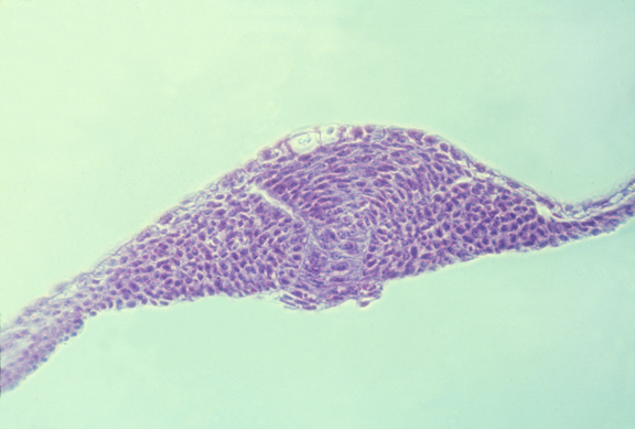
Fish embryo in the neurula stage:
---------------------------------------------------------------------------------------
Embryology of Frogs and Salamanders

Frogs and salamanders are both amphibians, and have similar embryos.
From the late 1800s until after the mid-1900s, most of the best embryological research was done using salamander & frog eggs.
Tissue culture was invented using cells from frog embryos, and clotted frog lymph as the substratum.
Advantages:
-
1) Large size of embryos (several millimeters in diameter)
2) Tolerate surgery very well (although large cells are very fragile)
(Early salamander embryos repair wounds in minutes or hours.)
Just keep the embryos is a very dilute salt (saline) solution, and you can tear loose organs and just squeeze them next to another area where you have removed the skin, and they quickly seal into position. Thousands of research papers have been published on such experiments.
The core of what we know, or think we know, about mechanisms of development is extrapolated from surgical research on amphibian eggs.
The kinds used most have been Xenopus (an African frog), and Amblystoma (includes the Spotted Salamanders of the eastern US) and also includes Axolotls (a semi-extinct Mexican Amblystoma) and also several kinds of Newts (a sub-group of salamanders)
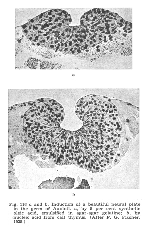
Hans Spemann and Hilde Prösholdt discovered embryonic induction of the neural tube by notochord mesoderm, using tissues from 2 newt species. (Spemann won a Nobel Prize specifically for discovering embryonic induction)
When signals from one part of an embryo stimulate differentiation of nearby cells into some different cell type than they otherwise would have become, this is called "Embryonic Induction"
Spemann's induction experiment
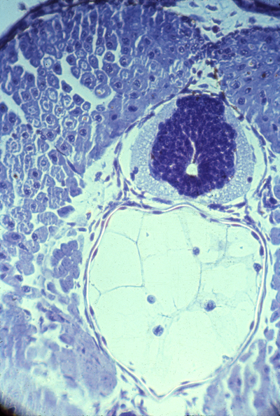
cross section of the neural tube and notochord
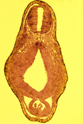
cross section of a developing tadpole
Links to videos: