Lecture notes for Friday, January 25, 2019
Shape Creation
Are there mechanical or chemical properties that can cause materials to gravitate toward becoming some certain particular three-dimensional shape?What properties tend to make materials become spherical?
What properties tend to make materials become tubular? (cylindrical)
If all tubular materials always have certain combinations of physical properties, then does that mean that all cells that have those physical properties will automatically rearrange to become tubular in shape?
Is that how genes cause anatomical shapes?
And is that how genes cause cells to rearrange into different shapes than they previously had?
Do genes cause cells to have properties (like certain contractilities of surfaces), with the properties causing the shapes?
How can you cause a sphere to become a tube (spontaneously)?
How could you revolutionize plastic surgery and orthopedic surgery using methods that cause cells to rearrange spontaneously into different geometric shapes?
Please visualize a hollow cylindrical pipe or tube, with a gas or liquid pushing outward.
The outward pressure will create tensions in whatever material the tube is made of (rubber, metal, cells, or whatever).
This tension will be exactly twice as strong in the direction going around the tube as compared with the direction parallel to the long axis of the tube.
-
How do we know?
Why should we believe?
Is there some way to prove that the ratio of directional tensions is two to one?
-
Read it in a book? (on the subject of "hoop stress" or "cylinder stress")
Measure tensions on surfaces of actual tubes?
Visualize a thought experiment.
The limitations on not too much wall thickness result from how much of the resistance is resistance to bending versus resistance to stretching.
(I am assuming that nearly all the important forces resist stretching, not bending even if the wall is fairly thick)
Please think about whether cylinders can be caused to form by means of their surface contractility becoming twice as strong in one direction as compared with the perpendicular direction.
***********************************************************************
Embryology of Frogs and Salamanders

Links to videos:
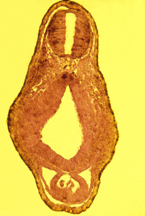
Cross section of a tadpole
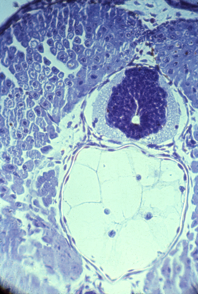
Amphibian neural tube and notochord
Experimental Embryology with Amphibians
Frogs and salamanders are both amphibians, and have similar embryos.From the late 1800s until after the mid-1900s, most of the best embryological research was done using salamander & frog eggs.
Tissue culture was invented using cells from frog embryos, and clotted frog lymph as the substratum.
Advantages:
-
1) Large size of embryos (several millimeters in diameter)
2) Tolerate surgery very well (although large cells are very fragile)
(Early salamander embryos repair wounds in minutes or hours.)
Just keep the embryos is a very dilute salt (saline) solution, and you can tear loose organs and just squeeze them next to another area where you have removed the skin, and they quickly seal into position. Thousands of research papers have been published on such experiments.
The core of what we know, or think we know, about mechanisms of development is extrapolated from surgical research on amphibian eggs.
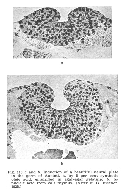
The kinds used most have been Xenopus (an African frog), and Amblystoma (includes the Spotted Salamanders of the eastern US) and also includes Axolotls (a semi-extinct Mexican Amblystoma) and also several kinds of Newts (a sub-group of salamanders).
Hans Spemann and Hilde Prösholdt discovered embryonic induction of the neural tube by notochord mesoderm, using tissues from 2 newt species. (Spemann won a Nobel Prize specifically for discovering embryonic induction.)
When signals from one part of an embryo stimulate differentiation of nearby cells into some different cell type than they otherwise would have become, this is called "Embryonic Induction."
Spemann's induction experiment
---------------------------------------------
"Embryonic Regulation"
Hans Driesch: discovery of "Regulative development" (in the 1890s). He separated the first 2, and the first 4 cells of starfish embryos, and observed their development into two half-size plutei, or 4 quarter-size. Conversely, you can push two one-cell stages together and sometimes they will develop into double-sized plutei.
This disproved the theory of different parts of embryos receiving different genes.
But even today scientists don't really understand the mechanism that changes the sizes of parts of embryos in proportion to the size of the whole embryo.
But please don't be misled by the word "regulation",
which can seem to imply some exterior control telling somebody what to do.
This kind of regulation is internal adjustment, to fix damage or overcome abnormality or effects of some external disturbance.
Whatever adjustments cause regulation of shape and adjustment of proportionality of sizes, sometimes require time to adjust.
"Regulative" development contrasts with "mosaic" development, in which cell fates are pre-determined, for example in the nematode Caenorhabditis elegans.
Driesch concluded that embryos are somewhat supernatural
in the sense of being controlled by something mind-like.
An even better example of size adjustment of parts in proportion to wholes is Dictyostelium formation of fruiting bodies, stalks, and length/width ratios of slime mold slugs.
If you cut a slug into three segments, all three will develop stalks and fruiting bodies. Even tiny pieces of a single slug will do this.
(Please understand that these "slugs" are masses of a kind of amoebae, & only happen to resemble real slugs, which are a kind of snail.)
Dictyostelium discoideum is the most-studied one of the "Cellular Slime Molds". In one stage of their life cycle, a culture consists of small amoeboid cells that move around, eating bacteria. When they run out of bacteria, they aggregate by chemotaxis, and form multicellular "slugs" that can crawl around. The slugs vary widely in size, depending on how many individual amoebae aggregated to form them. They then differentiate into stationary "fruiting bodies" that consist of a tapering stalk, with a lemon-shaped mass of spores at the top, and have the same proportions over a size range of at least 500.
This is even more than Driesch's revolutionary discovery that starfish embryos can "scale" over a sixteen-fold size range. (Incidentally, this most-studied species was discovered by a UNC undergraduate [Kenneth Raper] on some horse dung in Duke Forest.) For many years, the leading researcher on slime molds was Lindsay Olive, who was a professor in this department, and kindly provided many different species to use in laboratories of this course.
You can cut a Dictyostelium "slug" into 10 or 100 pieces, and the cells of each fragment will reorganize to form a normally-proportioned slug, and then a scale model stalk and spore mass, one one-hundredth the size the original "slug" would have formed. Echinoderm plutei can develop "scale models" over a volume ratio of 8 to 1 or sometimes 16 to one.
Scaling may have different mechanism in plutei than in slime mold fruiting bodies.
But I regard this as one of the 3 or 4 deepest questions in biology.
What does the ability "to scale" tell us about normal mechanisms that cause
organ formation?
That's the big question. There might be several answers, or one.
---------------------------------------------
Roux "hot needle" experiment (~1880s)
Wilhelm Roux did a famous experiment intended to confirm the theory of different parts of each embryo getting only certain genes. He poked red-hot needles into 4 or the first 8 cells or early frog embryos; or sometimes poked hot needles into 2 of the first 4 cells, or 1 of the first 2 cells.The idea was to kill or severely damage half the cells of an early embryo, with the expectation that the undamaged cells would continue and develop into the same tissues that they normally form, even though the "killed" cells failed to differentiate.
Textbooks use the verb "killed" to refer to the effect of the hot needle on embryonic frog cells. Actually, the original paper says the "killed" cells were still alive, and that after several hours they began to divide again.
Anyway, people interpreted the result as support for the idea of different genes being distributed unequally to different cells. (Nobody really understands the results of this experiment, but it has been repeated with the same results.)