Embryology of the Eye; Retino-tectal Connections
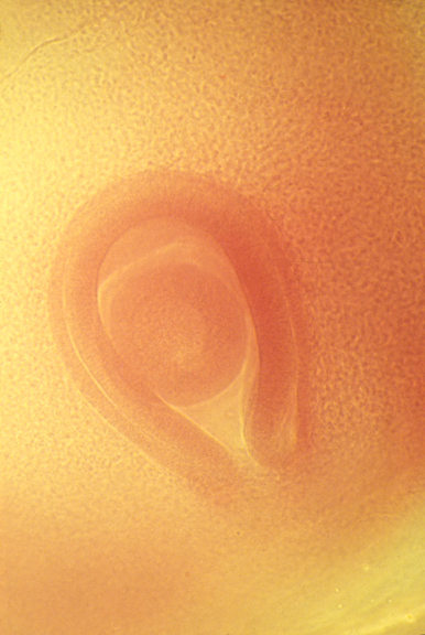
Embryonic chick eyeball, with retina surrounding it.
The groove is the location of the optic nerve.
Nerve fibers with growth cones at their tips are extended from the developing eye through the choroid fissure, and go to a specific area of the brain, the optic tectum. The locomotion of the growth cones is "amoeboid", of the same type that transports particles, as discussed in the lecture on January 24th.

Axons extending in tissue culture

Axons in tissue culture on a rubber substratum
Link to a video of axons extending in a cell culture
This was not shown in class, but please watch it.

Axons in brain, stained with silver
Nerve bodies in the brain are packed together closely. If you stained them all, you would see nothing but a solid dark color. The method used here randomly stains only a small percentage of the cells.
Horseradish peroxidase is an enzyme that catalyzes synthesis of a dark-colored product. It can be injected into the eye of an animal and used to track the nerves going from the eye to the brain.
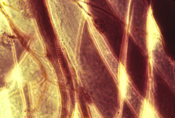
muscle cells with motor nerves innervating them
stained section of a whole developing brain

nerve ganglion cells in a collagen gel
"Low-magnification, dark-field microscopy view of living nerve axons crawling through a collagen gel, from an autonomic ganglion at the lower right corner toward the center and left. On the left side of this field, collagen has been strongly aligned (from lower left to upper center) by two clumps of strongly contractile mesenchymal cells (of which only the lower clump is in the field). The point is that the nerve axons are only slightly aligned. Live, unstained tissue." [from Harris, Albert K. (2014). Connective Tissue Development, in "Cell and Matrix Mechanics", eds. R.R. Kaunas and A. Zemel, Taylor and Francis Group, Boca Raton FL.]
Retino-Tectal Projection

Ganglion cells of the retina are the cells that actually connect to the brain.
They are on the innermost part of the neural retina, where light comes in through the rod and cone cells. (These are not the same thing as ganglia elsewhere in the body.) The rod and cone cells detect light and change the voltage polarization of the ganglion cells.
Each ganglion cell has an axon that crawls out of the eye to the brain.
A neural projection is where nerves in one place are connected into a pattern, then go to some other place and spread out into a map of the pattern from where they came, upside down and backwards.
If you cut the optic nerve of a salamander, growth cones will be re-formed in the correct pattern. Salamanders can also regenerate ganglion cells from the pigmented retina. Mammals can't do this.

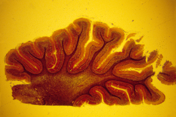
Cross-section of developing retina, showing neural retina at the left, pigmented retina, and some cartilage behind them

Cross-section of optic nerve on its way back to the brain
The "blind spot" in the retina has no nerve fibers sending messages back to the brain.

For many years people assumed that retino-tectal projections resulted from selective adhesiveness of retinal nerve cells. Experiments were done using radioactively labeled cells in an attempt to prove this (bottom of this figure; work by Steve Roth). Although dorsal retina cells did seem to stick preferentially to ventral optic tectum, there was never as much of a differential adhesion as was expected.

The real mechanism turns out to be gradients of selective detachment rather than adhesion, and involves proteins called Ephrins (in the optic tectum) and Ephrin receptors (Eph in this figure) in the retina. These are NOT diffusion gradients, but rather are gradients of relative sensitivity to inducing detachment. The molecules responsible are stuck in place. A nerve fiber with a lot of ephrin receptor doesn't need much ephrin to cause it to detach. A fiber with less ephrin receptor will go farther, and will detach only when there is a lot of ephrin.
(Think of running down a beach on very hot sand. If your feet are very sensitive, you won't run very far before it becomes more painful.)
Roth's experiment can be re-interpreted in terms of non-adhesiveness.