Specific examples of "Erroneous Papers", to practice writing reports about.
Please write a two page (or longer) report on your choice of either of the following two research papers. Both were published in very competitive journals, and their contents have not been publicly challenged.
Vasiokhin, V., C. Bauer, M. Yin, and E. Fuchs (2000). Directed actin polymerization is the driving force for epithelial cell-cell adhesion. Cell 100, 209-219. [link to journal]
Burton, K., and D.L. Taylor (1997). Traction forces of cytokinesis measured with optically modified elastic substrata. Nature 385, 450-454 [link to journal]
One paper reports new discoveries about cytoplasmic proteins inside cell protrusions that the authors assumed must be filopodia, poking out from the surfaces of tissue culture cells, as a result of increases in the calcium ion concentration in the culture media.
The authors of this paper did not know the literature well enough to realize that reduction in calcium ion concentrations in tissue culture media causes an increase in the flexibility of cell surfaces, so that membrane and cytoplasm get pulled out in long thin strands (called "retraction fibers") as the rest of each cell rounds up.
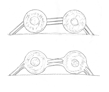
The same thing happens when cells undergo mitosis, after treatment with some proteolytic enzymes, and some antibodies. Observation of retraction fibers was an important part of the discovery of focal adhesions, the existence of which was once controversial, but is now accepted. [See Harris, A K. (1973). Location of cellular adhesions to solid substrata. Dev. Biol. 35: 97-114.] Retraction fibers usually are so narrow (tenth of a micrometer) that they cannot be resolved by light microscopes. They are frequently seen in scanning electron microscope pictures, and interpreted as filopodia. Often retraction fibers form along the rear margin of crawling cells; when these are seen in scanning electron microscopy, they are almost always interpreted as filopodia, and their location is thought to be the front of the cell.
Following mitosis, filopodia get re-inflated with cytoplasm. Increasing the calcium concentration also causes re-inflation of retraction fibers with cytoplasm. As the diameter of retraction fibers increases, they become thick enough to be resolved by light microscopy. Sloppy researchers misinterpret the thicker parts as being the tip ends of filopodia, being pushed out from the cells. Really, they are more like strands of Mozzarella cheese being pulled out in long strands as a pizza is separated into pieces. But they get misinterpreted as if they were long spines on a sea urchin. Notice the illustrations in which "filopodia" point directly at each other.
Please do not confuse honest mistakes of this kind as if it were research fraud. Nevertheless, the effect is as bad or worse. In my opinion, this kind of sloppy research is why there is still no cure for cancer or many other diseases. It uses up tens of millions of dollars of grant money, perpetuates erroneous paradigms, but rarely results in either honest retraction or punishment.
The second paper, by Burton and Taylor is much better, and may or may not be erroneous. They improved a previously invented experimental method, improved its quantitative accuracy, and used it to test the locations and directions of forces exerted by cell surfaces at different stages of mitosis. One of the photographs was published on the front cover of Nature, and seems to show a localization of contractile forces parallel to the mitotic furrow. Perhaps it does. But a tricky fact about these wrinkles is that they form perpendicular to the main direction of force, and also parallel to the direction of force. The authors were aware of this, and did their best to distinguish directions of forces. They may have been 90 degrees off.
Researchers often use gels to detect directions and strengths of cellular forces. Gels were used for many years before rubber layers. But unti the rubber method, the dogma was that gels shrank in response to biochemical changes caused by cell metabolism. Because silicone rubber is chemically inert, its distortion could not be biochemical, and the wrinkles were visible in still photographs, and didn't need movie films to make distortion visible. You wouldn't believe how controversial this issue was, until settled by the rubber sheet method.
Incidentally, when tension is applied to a gel along one axis, this produces two distortions, one parallel to the tension and the other exactly perpendicular to it. Researchers who use gels to study cell forces seldom realize this. They think rubber sheets are inferior to gels, because you get two perpendicular sets of wrinkles. But at least you can see both of them. In gels, two perpendicular distortions also occur, and get confused with each other.
It is a good question why very fundamental issues remain unsolved. Reading these papers can help you understand why. The truth can hide in plain sight.